Iron »
PDB 3na1-3nmj »
3na1 »
Iron in PDB 3na1: Crystal Structure of Human CYP11A1 in Complex with 20- Hydroxycholesterol
Enzymatic activity of Crystal Structure of Human CYP11A1 in Complex with 20- Hydroxycholesterol
All present enzymatic activity of Crystal Structure of Human CYP11A1 in Complex with 20- Hydroxycholesterol:
1.14.15.6;
1.14.15.6;
Protein crystallography data
The structure of Crystal Structure of Human CYP11A1 in Complex with 20- Hydroxycholesterol, PDB code: 3na1
was solved by
N.V.Strushkevich,
F.Mackenzie,
W.Tempel,
A.Botchkarev,
C.H.Arrowsmith,
A.M.Edwards,
C.Bountra,
J.U.Weigelt,
H.Park,
Structural Genomicsconsortium (Sgc),
with X-Ray Crystallography technique. A brief refinement statistics is given in the table below:
| Resolution Low / High (Å) | 50.00 / 2.25 |
| Space group | P 1 21 1 |
| Cell size a, b, c (Å), α, β, γ (°) | 82.810, 115.149, 85.727, 90.00, 101.45, 90.00 |
| R / Rfree (%) | 19.3 / 23.2 |
Iron Binding Sites:
The binding sites of Iron atom in the Crystal Structure of Human CYP11A1 in Complex with 20- Hydroxycholesterol
(pdb code 3na1). This binding sites where shown within
5.0 Angstroms radius around Iron atom.
In total 6 binding sites of Iron where determined in the Crystal Structure of Human CYP11A1 in Complex with 20- Hydroxycholesterol, PDB code: 3na1:
Jump to Iron binding site number: 1; 2; 3; 4; 5; 6;
In total 6 binding sites of Iron where determined in the Crystal Structure of Human CYP11A1 in Complex with 20- Hydroxycholesterol, PDB code: 3na1:
Jump to Iron binding site number: 1; 2; 3; 4; 5; 6;
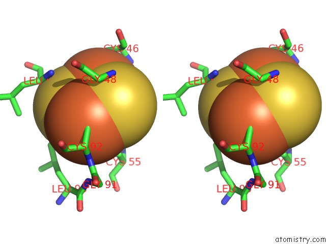
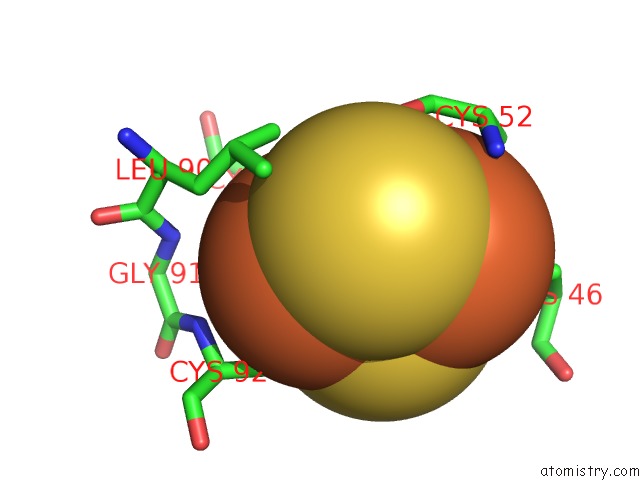
Iron binding site 1 out of 6 in 3na1
Go back to
Iron binding site 1 out
of 6 in the Crystal Structure of Human CYP11A1 in Complex with 20- Hydroxycholesterol

Mono view
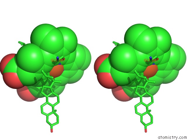
Stereo pair view
Mono view
Stereo pair view
A full contact list of Iron with other atoms in the Fe binding
site number 1 of Crystal Structure of Human CYP11A1 in Complex with 20- Hydroxycholesterol within 5.0Å range:
|
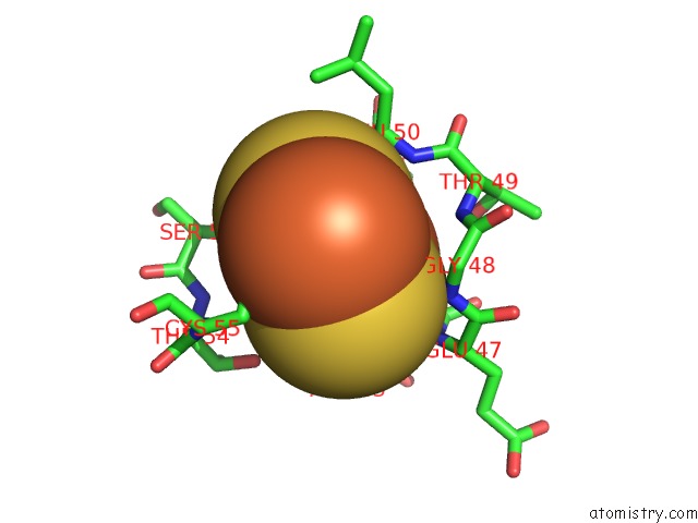
Iron binding site 2 out of 6 in 3na1
Go back to
Iron binding site 2 out
of 6 in the Crystal Structure of Human CYP11A1 in Complex with 20- Hydroxycholesterol

Mono view

Stereo pair view
Mono view
Stereo pair view
A full contact list of Iron with other atoms in the Fe binding
site number 2 of Crystal Structure of Human CYP11A1 in Complex with 20- Hydroxycholesterol within 5.0Å range:
|
Iron binding site 3 out of 6 in 3na1
Go back to
Iron binding site 3 out
of 6 in the Crystal Structure of Human CYP11A1 in Complex with 20- Hydroxycholesterol
Mono view
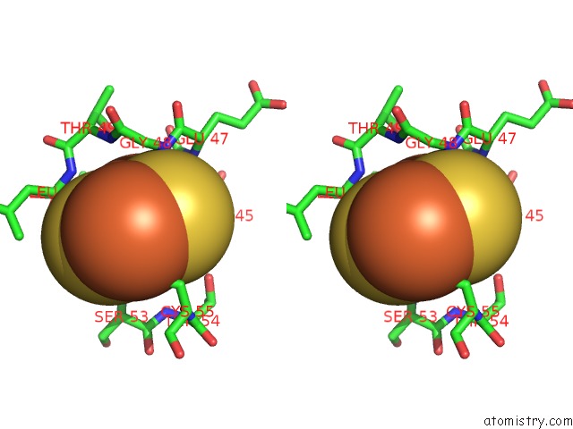
Stereo pair view
Mono view
Stereo pair view
A full contact list of Iron with other atoms in the Fe binding
site number 3 of Crystal Structure of Human CYP11A1 in Complex with 20- Hydroxycholesterol within 5.0Å range:
|
Iron binding site 4 out of 6 in 3na1
Go back to
Iron binding site 4 out
of 6 in the Crystal Structure of Human CYP11A1 in Complex with 20- Hydroxycholesterol
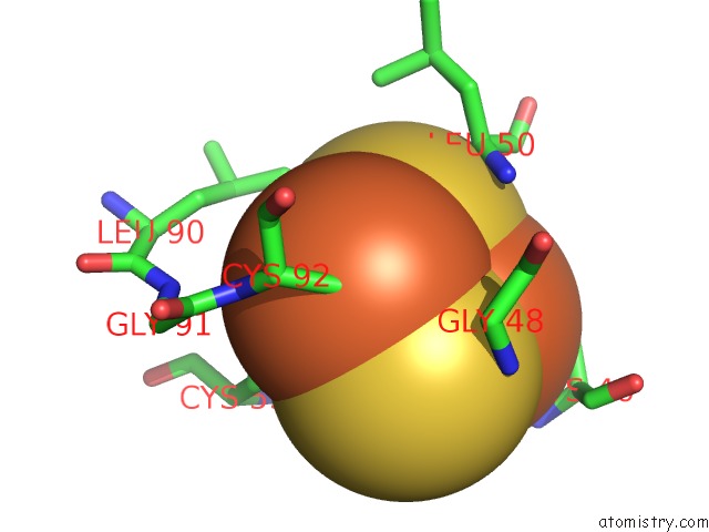
Mono view
Stereo pair view
Mono view
Stereo pair view
A full contact list of Iron with other atoms in the Fe binding
site number 4 of Crystal Structure of Human CYP11A1 in Complex with 20- Hydroxycholesterol within 5.0Å range:
|
Iron binding site 5 out of 6 in 3na1
Go back to
Iron binding site 5 out
of 6 in the Crystal Structure of Human CYP11A1 in Complex with 20- Hydroxycholesterol

Mono view

Stereo pair view
Mono view
Stereo pair view
A full contact list of Iron with other atoms in the Fe binding
site number 5 of Crystal Structure of Human CYP11A1 in Complex with 20- Hydroxycholesterol within 5.0Å range:
|
Iron binding site 6 out of 6 in 3na1
Go back to
Iron binding site 6 out
of 6 in the Crystal Structure of Human CYP11A1 in Complex with 20- Hydroxycholesterol
Mono view

Stereo pair view
Mono view
Stereo pair view
A full contact list of Iron with other atoms in the Fe binding
site number 6 of Crystal Structure of Human CYP11A1 in Complex with 20- Hydroxycholesterol within 5.0Å range:
|
Reference:
N.Strushkevich,
F.Mackenzie,
T.Cherkesova,
I.Grabovec,
S.Usanov,
H.W.Park.
Structural Basis For Pregnenolone Biosynthesis By the Mitochondrial Monooxygenase System. Proc.Natl.Acad.Sci.Usa V. 108 10139 2011.
ISSN: ISSN 0027-8424
PubMed: 21636783
DOI: 10.1073/PNAS.1019441108
Page generated: Sun Aug 4 16:24:57 2024
ISSN: ISSN 0027-8424
PubMed: 21636783
DOI: 10.1073/PNAS.1019441108
Last articles
F in 4QBJF in 4Q9R
F in 4Q8X
F in 4Q83
F in 4Q87
F in 4Q7B
F in 4Q0E
F in 4Q1E
F in 4Q3K
F in 4Q1D