Iron »
PDB 3na1-3nmj »
3nbs »
Iron in PDB 3nbs: Crystal Structure of Dimeric Cytochrome C From Horse Heart
Protein crystallography data
The structure of Crystal Structure of Dimeric Cytochrome C From Horse Heart, PDB code: 3nbs
was solved by
M.Taketa,
H.Komori,
S.Hirota,
Y.Higuchi,
with X-Ray Crystallography technique. A brief refinement statistics is given in the table below:
| Resolution Low / High (Å) | 19.58 / 2.20 |
| Space group | P 1 |
| Cell size a, b, c (Å), α, β, γ (°) | 41.753, 56.305, 60.815, 66.28, 89.94, 73.66 |
| R / Rfree (%) | 21.5 / 27.3 |
Iron Binding Sites:
The binding sites of Iron atom in the Crystal Structure of Dimeric Cytochrome C From Horse Heart
(pdb code 3nbs). This binding sites where shown within
5.0 Angstroms radius around Iron atom.
In total 4 binding sites of Iron where determined in the Crystal Structure of Dimeric Cytochrome C From Horse Heart, PDB code: 3nbs:
Jump to Iron binding site number: 1; 2; 3; 4;
In total 4 binding sites of Iron where determined in the Crystal Structure of Dimeric Cytochrome C From Horse Heart, PDB code: 3nbs:
Jump to Iron binding site number: 1; 2; 3; 4;
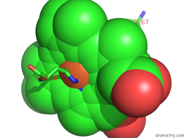
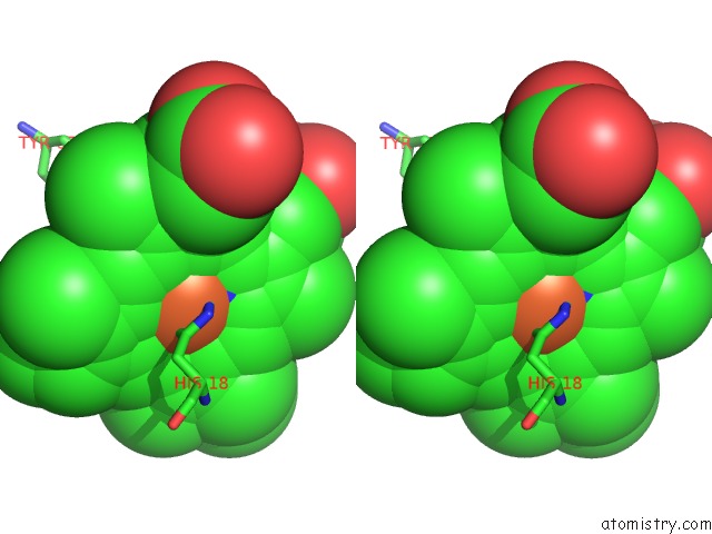
Iron binding site 1 out of 4 in 3nbs
Go back to
Iron binding site 1 out
of 4 in the Crystal Structure of Dimeric Cytochrome C From Horse Heart
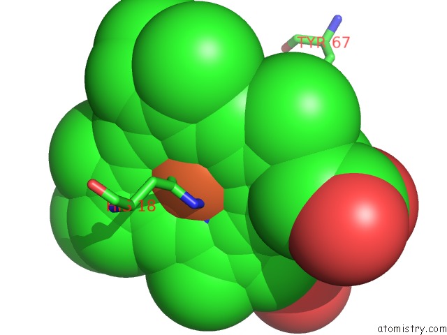
Mono view
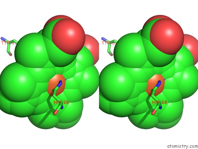
Stereo pair view
Mono view
Stereo pair view
A full contact list of Iron with other atoms in the Fe binding
site number 1 of Crystal Structure of Dimeric Cytochrome C From Horse Heart within 5.0Å range:
|
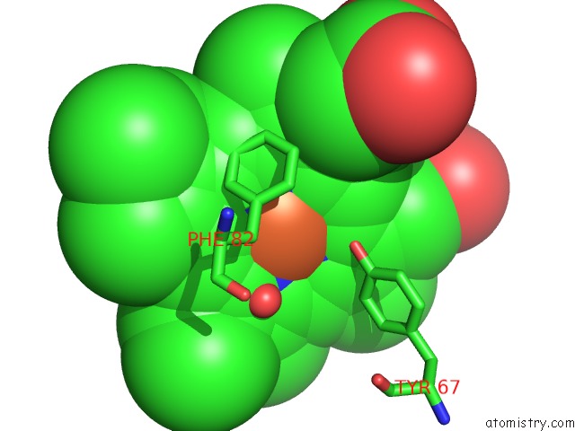
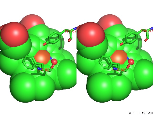
Iron binding site 2 out of 4 in 3nbs
Go back to
Iron binding site 2 out
of 4 in the Crystal Structure of Dimeric Cytochrome C From Horse Heart
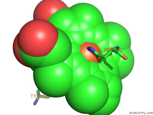
Mono view
Stereo pair view
Mono view
Stereo pair view
A full contact list of Iron with other atoms in the Fe binding
site number 2 of Crystal Structure of Dimeric Cytochrome C From Horse Heart within 5.0Å range:
|
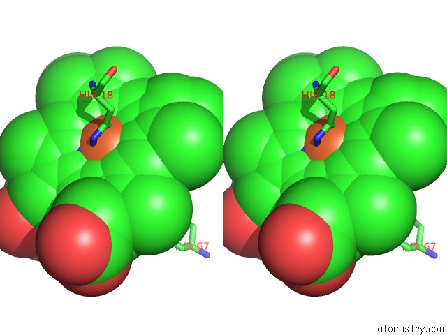
Iron binding site 3 out of 4 in 3nbs
Go back to
Iron binding site 3 out
of 4 in the Crystal Structure of Dimeric Cytochrome C From Horse Heart
Mono view
Stereo pair view
Mono view
Stereo pair view
A full contact list of Iron with other atoms in the Fe binding
site number 3 of Crystal Structure of Dimeric Cytochrome C From Horse Heart within 5.0Å range:
|
Iron binding site 4 out of 4 in 3nbs
Go back to
Iron binding site 4 out
of 4 in the Crystal Structure of Dimeric Cytochrome C From Horse Heart
Mono view
Stereo pair view
Mono view
Stereo pair view
A full contact list of Iron with other atoms in the Fe binding
site number 4 of Crystal Structure of Dimeric Cytochrome C From Horse Heart within 5.0Å range:
|
Reference:
S.Hirota,
Y.Hattori,
S.Nagao,
M.Taketa,
H.Komori,
H.Kamikubo,
Z.Wang,
I.Takahashi,
S.Negi,
Y.Sugiura,
M.Kataoka,
Y.Higuchi.
Cytochrome C Polymerization By Successive Domain Swapping at the C-Terminal Helix Proc.Natl.Acad.Sci.Usa V. 107 12854 2010.
ISSN: ISSN 0027-8424
PubMed: 20615990
DOI: 10.1073/PNAS.1001839107
Page generated: Sun Aug 4 16:24:56 2024
ISSN: ISSN 0027-8424
PubMed: 20615990
DOI: 10.1073/PNAS.1001839107
Last articles
Zn in 9J0NZn in 9J0O
Zn in 9J0P
Zn in 9FJX
Zn in 9EKB
Zn in 9C0F
Zn in 9CAH
Zn in 9CH0
Zn in 9CH3
Zn in 9CH1