Iron »
PDB 3na1-3nmj »
3ng6 »
Iron in PDB 3ng6: The Crystal Structure of Hemoglobin I From Trematomus Newnesi in Deoxygenated State Obtained Through An Oxidation/Reduction Cycle in Which Potassium Hexacyanoferrate and Sodium Dithionite Were Alternatively Added
Protein crystallography data
The structure of The Crystal Structure of Hemoglobin I From Trematomus Newnesi in Deoxygenated State Obtained Through An Oxidation/Reduction Cycle in Which Potassium Hexacyanoferrate and Sodium Dithionite Were Alternatively Added, PDB code: 3ng6
was solved by
A.Vergara,
L.Vitagliano,
A.Merlino,
F.Sica,
K.Marino,
L.Mazzarella,
with X-Ray Crystallography technique. A brief refinement statistics is given in the table below:
| Resolution Low / High (Å) | 26.60 / 2.20 |
| Space group | P 41 |
| Cell size a, b, c (Å), α, β, γ (°) | 61.882, 61.882, 187.014, 90.00, 90.00, 90.00 |
| R / Rfree (%) | 19.5 / 24.7 |
Iron Binding Sites:
The binding sites of Iron atom in the The Crystal Structure of Hemoglobin I From Trematomus Newnesi in Deoxygenated State Obtained Through An Oxidation/Reduction Cycle in Which Potassium Hexacyanoferrate and Sodium Dithionite Were Alternatively Added
(pdb code 3ng6). This binding sites where shown within
5.0 Angstroms radius around Iron atom.
In total 4 binding sites of Iron where determined in the The Crystal Structure of Hemoglobin I From Trematomus Newnesi in Deoxygenated State Obtained Through An Oxidation/Reduction Cycle in Which Potassium Hexacyanoferrate and Sodium Dithionite Were Alternatively Added, PDB code: 3ng6:
Jump to Iron binding site number: 1; 2; 3; 4;
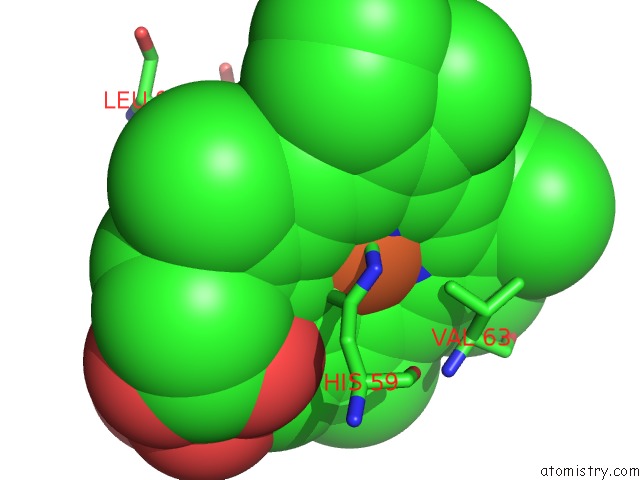
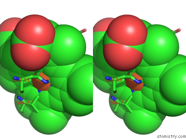
Iron binding site 1 out of 4 in 3ng6
Go back to
Iron binding site 1 out
of 4 in the The Crystal Structure of Hemoglobin I From Trematomus Newnesi in Deoxygenated State Obtained Through An Oxidation/Reduction Cycle in Which Potassium Hexacyanoferrate and Sodium Dithionite Were Alternatively Added
Mono view
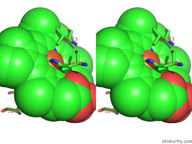
Stereo pair view
Mono view
Stereo pair view
A full contact list of Iron with other atoms in the Fe binding
site number 1 of The Crystal Structure of Hemoglobin I From Trematomus Newnesi in Deoxygenated State Obtained Through An Oxidation/Reduction Cycle in Which Potassium Hexacyanoferrate and Sodium Dithionite Were Alternatively Added within 5.0Å range:
|
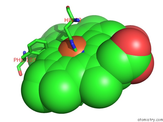
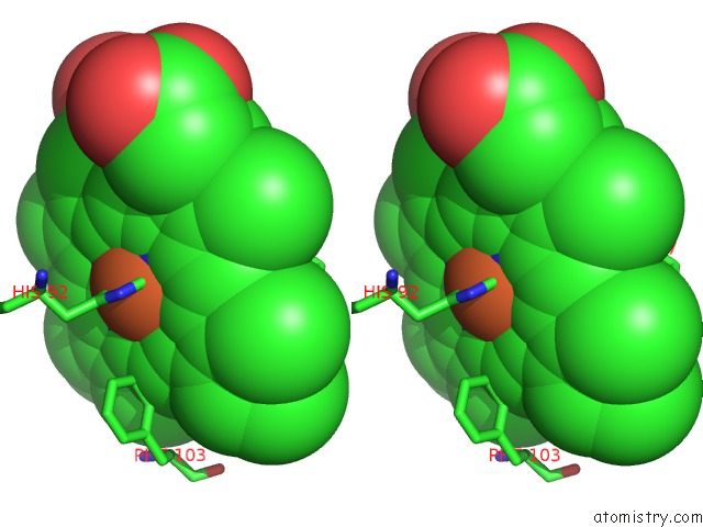
Iron binding site 2 out of 4 in 3ng6
Go back to
Iron binding site 2 out
of 4 in the The Crystal Structure of Hemoglobin I From Trematomus Newnesi in Deoxygenated State Obtained Through An Oxidation/Reduction Cycle in Which Potassium Hexacyanoferrate and Sodium Dithionite Were Alternatively Added

Mono view
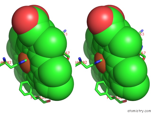
Stereo pair view
Mono view
Stereo pair view
A full contact list of Iron with other atoms in the Fe binding
site number 2 of The Crystal Structure of Hemoglobin I From Trematomus Newnesi in Deoxygenated State Obtained Through An Oxidation/Reduction Cycle in Which Potassium Hexacyanoferrate and Sodium Dithionite Were Alternatively Added within 5.0Å range:
|
Iron binding site 3 out of 4 in 3ng6
Go back to
Iron binding site 3 out
of 4 in the The Crystal Structure of Hemoglobin I From Trematomus Newnesi in Deoxygenated State Obtained Through An Oxidation/Reduction Cycle in Which Potassium Hexacyanoferrate and Sodium Dithionite Were Alternatively Added
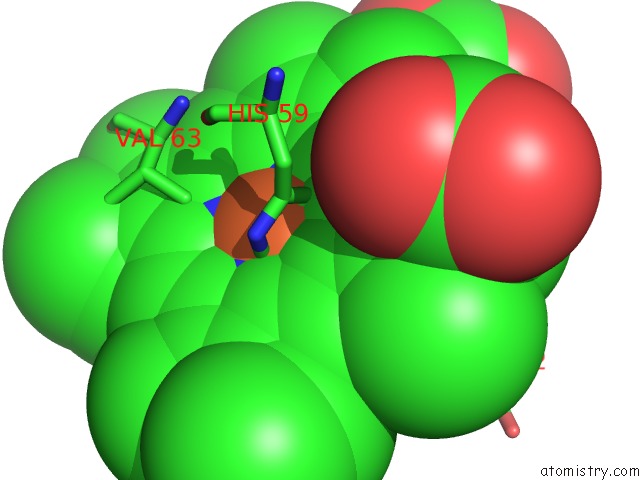
Mono view
Stereo pair view
Mono view
Stereo pair view
A full contact list of Iron with other atoms in the Fe binding
site number 3 of The Crystal Structure of Hemoglobin I From Trematomus Newnesi in Deoxygenated State Obtained Through An Oxidation/Reduction Cycle in Which Potassium Hexacyanoferrate and Sodium Dithionite Were Alternatively Added within 5.0Å range:
|
Iron binding site 4 out of 4 in 3ng6
Go back to
Iron binding site 4 out
of 4 in the The Crystal Structure of Hemoglobin I From Trematomus Newnesi in Deoxygenated State Obtained Through An Oxidation/Reduction Cycle in Which Potassium Hexacyanoferrate and Sodium Dithionite Were Alternatively Added
Mono view
Stereo pair view
Mono view
Stereo pair view
A full contact list of Iron with other atoms in the Fe binding
site number 4 of The Crystal Structure of Hemoglobin I From Trematomus Newnesi in Deoxygenated State Obtained Through An Oxidation/Reduction Cycle in Which Potassium Hexacyanoferrate and Sodium Dithionite Were Alternatively Added within 5.0Å range:
|
Reference:
A.Vergara,
L.Vitagliano,
A.Merlino,
F.Sica,
K.Marino,
C.Verde,
G.Di Prisco,
L.Mazzarella.
An Order-Disorder Transition Plays A Role in Switching Off the Root Effect in Fish Hemoglobins. J.Biol.Chem. V. 285 32568 2010.
ISSN: ISSN 0021-9258
PubMed: 20610398
DOI: 10.1074/JBC.M110.143537
Page generated: Sun Aug 4 16:27:22 2024
ISSN: ISSN 0021-9258
PubMed: 20610398
DOI: 10.1074/JBC.M110.143537
Last articles
Zn in 9MJ5Zn in 9HNW
Zn in 9G0L
Zn in 9FNE
Zn in 9DZN
Zn in 9E0I
Zn in 9D32
Zn in 9DAK
Zn in 8ZXC
Zn in 8ZUF