Iron »
PDB 3na1-3nmj »
3njz »
Iron in PDB 3njz: Crystal Structure of Salicylate 1,2-Dioxygenase From Pseudoaminobacter Salicylatoxidans Adducts with Salicylate
Enzymatic activity of Crystal Structure of Salicylate 1,2-Dioxygenase From Pseudoaminobacter Salicylatoxidans Adducts with Salicylate
All present enzymatic activity of Crystal Structure of Salicylate 1,2-Dioxygenase From Pseudoaminobacter Salicylatoxidans Adducts with Salicylate:
1.13.11.4;
1.13.11.4;
Protein crystallography data
The structure of Crystal Structure of Salicylate 1,2-Dioxygenase From Pseudoaminobacter Salicylatoxidans Adducts with Salicylate, PDB code: 3njz
was solved by
M.Ferraroni,
F.Briganti,
I.Matera,
with X-Ray Crystallography technique. A brief refinement statistics is given in the table below:
| Resolution Low / High (Å) | 30.00 / 2.10 |
| Space group | I 2 2 2 |
| Cell size a, b, c (Å), α, β, γ (°) | 76.652, 86.961, 166.465, 90.00, 90.00, 90.00 |
| R / Rfree (%) | 19.1 / 23.1 |
Iron Binding Sites:
The binding sites of Iron atom in the Crystal Structure of Salicylate 1,2-Dioxygenase From Pseudoaminobacter Salicylatoxidans Adducts with Salicylate
(pdb code 3njz). This binding sites where shown within
5.0 Angstroms radius around Iron atom.
In total only one binding site of Iron was determined in the Crystal Structure of Salicylate 1,2-Dioxygenase From Pseudoaminobacter Salicylatoxidans Adducts with Salicylate, PDB code: 3njz:
In total only one binding site of Iron was determined in the Crystal Structure of Salicylate 1,2-Dioxygenase From Pseudoaminobacter Salicylatoxidans Adducts with Salicylate, PDB code: 3njz:
Iron binding site 1 out of 1 in 3njz
Go back to
Iron binding site 1 out
of 1 in the Crystal Structure of Salicylate 1,2-Dioxygenase From Pseudoaminobacter Salicylatoxidans Adducts with Salicylate
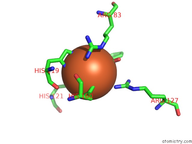
Mono view
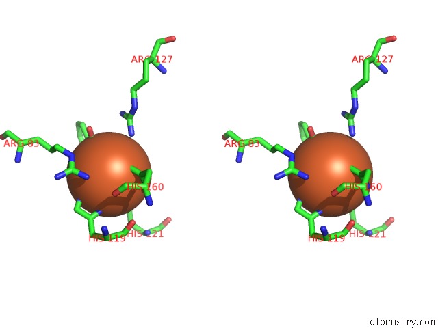
Stereo pair view
Mono view
Stereo pair view
A full contact list of Iron with other atoms in the Fe binding
site number 1 of Crystal Structure of Salicylate 1,2-Dioxygenase From Pseudoaminobacter Salicylatoxidans Adducts with Salicylate within 5.0Å range:
|
Reference:
M.Ferraroni,
I.Matera,
L.Steimer,
S.Burger,
A.Scozzafava,
A.Stolz,
F.Briganti.
Crystal Structures of Salicylate 1,2-Dioxygenase-Substrates Adducts: A Step Towards the Comprehension of the Structural Basis For Substrate Selection in Class III Ring Cleaving Dioxygenases. J.Struct.Biol. V. 177 431 2012.
ISSN: ISSN 1047-8477
PubMed: 22155290
DOI: 10.1016/J.JSB.2011.11.026
Page generated: Sun Aug 4 16:27:39 2024
ISSN: ISSN 1047-8477
PubMed: 22155290
DOI: 10.1016/J.JSB.2011.11.026
Last articles
Zn in 9J0NZn in 9J0O
Zn in 9J0P
Zn in 9FJX
Zn in 9EKB
Zn in 9C0F
Zn in 9CAH
Zn in 9CH0
Zn in 9CH3
Zn in 9CH1