Iron »
PDB 3na1-3nmj »
3nmj »
Iron in PDB 3nmj: Crystal Structure of A Nickel Mediated Dimer For the Phenanthroline- Modified Cytochrome CB562 Variant, Mbp-PHEN2
Protein crystallography data
The structure of Crystal Structure of A Nickel Mediated Dimer For the Phenanthroline- Modified Cytochrome CB562 Variant, Mbp-PHEN2, PDB code: 3nmj
was solved by
R.J.Radford,
F.A.Tezcan,
with X-Ray Crystallography technique. A brief refinement statistics is given in the table below:
| Resolution Low / High (Å) | 85.99 / 3.10 |
| Space group | P 65 |
| Cell size a, b, c (Å), α, β, γ (°) | 99.291, 99.291, 109.317, 90.00, 90.00, 120.00 |
| R / Rfree (%) | 19.3 / 23.8 |
Other elements in 3nmj:
The structure of Crystal Structure of A Nickel Mediated Dimer For the Phenanthroline- Modified Cytochrome CB562 Variant, Mbp-PHEN2 also contains other interesting chemical elements:
| Nickel | (Ni) | 2 atoms |
Iron Binding Sites:
The binding sites of Iron atom in the Crystal Structure of A Nickel Mediated Dimer For the Phenanthroline- Modified Cytochrome CB562 Variant, Mbp-PHEN2
(pdb code 3nmj). This binding sites where shown within
5.0 Angstroms radius around Iron atom.
In total 4 binding sites of Iron where determined in the Crystal Structure of A Nickel Mediated Dimer For the Phenanthroline- Modified Cytochrome CB562 Variant, Mbp-PHEN2, PDB code: 3nmj:
Jump to Iron binding site number: 1; 2; 3; 4;
In total 4 binding sites of Iron where determined in the Crystal Structure of A Nickel Mediated Dimer For the Phenanthroline- Modified Cytochrome CB562 Variant, Mbp-PHEN2, PDB code: 3nmj:
Jump to Iron binding site number: 1; 2; 3; 4;
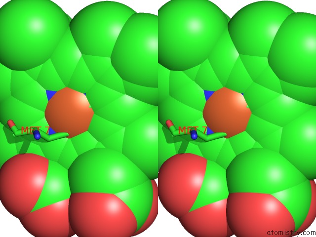
Iron binding site 1 out of 4 in 3nmj
Go back to
Iron binding site 1 out
of 4 in the Crystal Structure of A Nickel Mediated Dimer For the Phenanthroline- Modified Cytochrome CB562 Variant, Mbp-PHEN2

Mono view
Stereo pair view
Mono view
Stereo pair view
A full contact list of Iron with other atoms in the Fe binding
site number 1 of Crystal Structure of A Nickel Mediated Dimer For the Phenanthroline- Modified Cytochrome CB562 Variant, Mbp-PHEN2 within 5.0Å range:
|
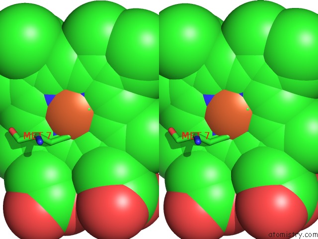
Iron binding site 2 out of 4 in 3nmj
Go back to
Iron binding site 2 out
of 4 in the Crystal Structure of A Nickel Mediated Dimer For the Phenanthroline- Modified Cytochrome CB562 Variant, Mbp-PHEN2

Mono view

Stereo pair view
Mono view
Stereo pair view
A full contact list of Iron with other atoms in the Fe binding
site number 2 of Crystal Structure of A Nickel Mediated Dimer For the Phenanthroline- Modified Cytochrome CB562 Variant, Mbp-PHEN2 within 5.0Å range:
|
Iron binding site 3 out of 4 in 3nmj
Go back to
Iron binding site 3 out
of 4 in the Crystal Structure of A Nickel Mediated Dimer For the Phenanthroline- Modified Cytochrome CB562 Variant, Mbp-PHEN2

Mono view

Stereo pair view
Mono view
Stereo pair view
A full contact list of Iron with other atoms in the Fe binding
site number 3 of Crystal Structure of A Nickel Mediated Dimer For the Phenanthroline- Modified Cytochrome CB562 Variant, Mbp-PHEN2 within 5.0Å range:
|
Iron binding site 4 out of 4 in 3nmj
Go back to
Iron binding site 4 out
of 4 in the Crystal Structure of A Nickel Mediated Dimer For the Phenanthroline- Modified Cytochrome CB562 Variant, Mbp-PHEN2

Mono view
Stereo pair view
Mono view
Stereo pair view
A full contact list of Iron with other atoms in the Fe binding
site number 4 of Crystal Structure of A Nickel Mediated Dimer For the Phenanthroline- Modified Cytochrome CB562 Variant, Mbp-PHEN2 within 5.0Å range:
|
Reference:
R.J.Radford,
M.Lawrenz,
P.C.Nguyen,
J.A.Mccammon,
F.A.Tezcan.
Porous Protein Frameworks with Unsaturated Metal Centers in Sterically Encumbered Coordination Sites. Chem.Commun.(Camb.) V. 47 313 2011.
ISSN: ISSN 1359-7345
PubMed: 20740227
DOI: 10.1039/C0CC02168G
Page generated: Tue Aug 5 04:49:13 2025
ISSN: ISSN 1359-7345
PubMed: 20740227
DOI: 10.1039/C0CC02168G
Last articles
Fe in 3VM1Fe in 3VM0
Fe in 3VM9
Fe in 3VM4
Fe in 3VLY
Fe in 3VLX
Fe in 3VLZ
Fe in 3VKT
Fe in 3VLL
Fe in 3VLM