Iron in PDB 3ns1: Crystal Structure of Bovine Xanthine Oxidase in Complex with 6- Mercaptopurine
Enzymatic activity of Crystal Structure of Bovine Xanthine Oxidase in Complex with 6- Mercaptopurine
All present enzymatic activity of Crystal Structure of Bovine Xanthine Oxidase in Complex with 6- Mercaptopurine:
1.17.1.4;
1.17.3.2;
Protein crystallography data
The structure of Crystal Structure of Bovine Xanthine Oxidase in Complex with 6- Mercaptopurine, PDB code: 3ns1
was solved by
H.Cao,
J.M.Pauff,
R.Hille,
with X-Ray Crystallography technique. A brief refinement statistics is given in the table below:
Resolution Low / High (Å)
|
45.50 /
2.60
|
Space group
|
P 1 21 1
|
Cell size a, b, c (Å), α, β, γ (°)
|
134.636,
73.928,
140.353,
90.00,
97.01,
90.00
|
R / Rfree (%)
|
21.7 /
26.9
|
Other elements in 3ns1:
The structure of Crystal Structure of Bovine Xanthine Oxidase in Complex with 6- Mercaptopurine also contains other interesting chemical elements:
Iron Binding Sites:
The binding sites of Iron atom in the Crystal Structure of Bovine Xanthine Oxidase in Complex with 6- Mercaptopurine
(pdb code 3ns1). This binding sites where shown within
5.0 Angstroms radius around Iron atom.
In total 8 binding sites of Iron where determined in the
Crystal Structure of Bovine Xanthine Oxidase in Complex with 6- Mercaptopurine, PDB code: 3ns1:
Jump to Iron binding site number:
1;
2;
3;
4;
5;
6;
7;
8;
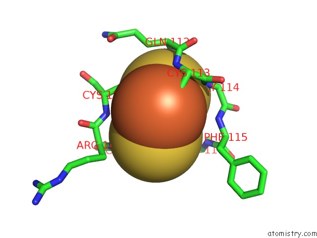
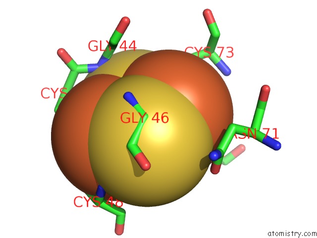
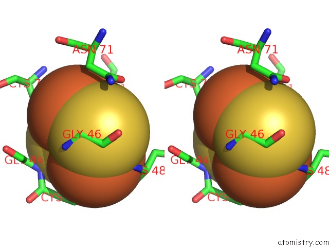
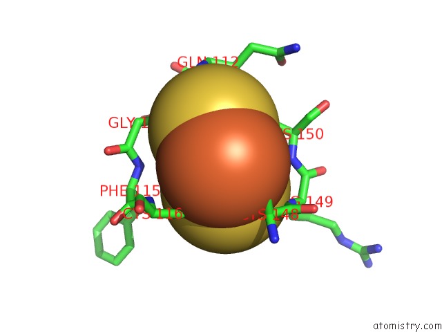
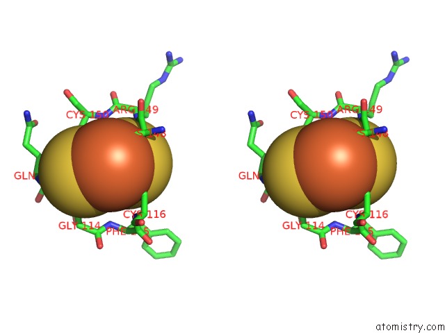
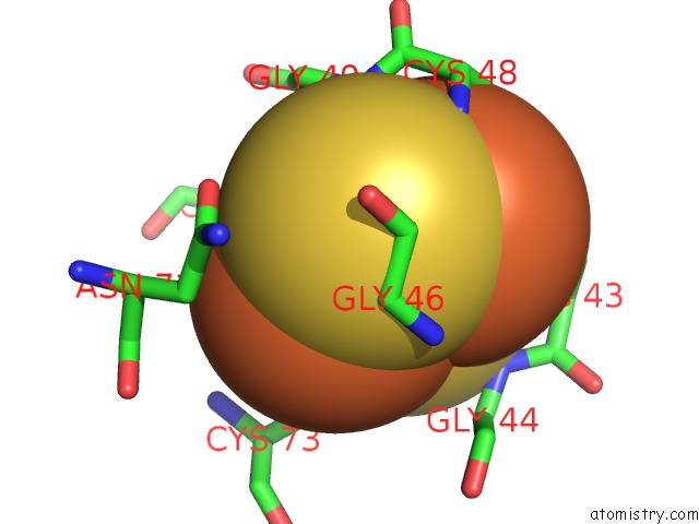
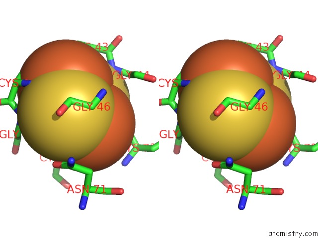
Iron binding site 1 out
of 8 in 3ns1
Go back to
Iron Binding Sites List in 3ns1
Iron binding site 1 out
of 8 in the Crystal Structure of Bovine Xanthine Oxidase in Complex with 6- Mercaptopurine
 Mono view
 Stereo pair view
|
A full contact list of Iron with other atoms in the Fe binding
site number 1 of Crystal Structure of Bovine Xanthine Oxidase in Complex with 6- Mercaptopurine within 5.0Å range:
probe
|
atom
|
residue
|
distance (Å)
|
B
|
Occ
|
A:Fe601
b:17.1
occ:1.00
|
FE1
|
A:FES601
|
0.0
|
17.1
|
1.0
|
S1
|
A:FES601
|
2.2
|
12.6
|
1.0
|
S2
|
A:FES601
|
2.2
|
12.6
|
1.0
|
SG
|
A:CYS150
|
2.4
|
17.6
|
1.0
|
SG
|
A:CYS113
|
2.4
|
15.8
|
1.0
|
FE2
|
A:FES601
|
2.9
|
14.1
|
1.0
|
CB
|
A:CYS150
|
3.3
|
20.5
|
1.0
|
CB
|
A:CYS113
|
3.4
|
16.1
|
1.0
|
N
|
A:CYS113
|
3.7
|
16.6
|
1.0
|
N
|
A:GLY114
|
4.0
|
14.8
|
1.0
|
CA
|
A:CYS113
|
4.0
|
15.6
|
1.0
|
N
|
A:CYS150
|
4.0
|
19.7
|
1.0
|
CA
|
A:CYS150
|
4.3
|
19.7
|
1.0
|
C
|
A:CYS113
|
4.3
|
15.7
|
1.0
|
SG
|
A:CYS148
|
4.5
|
15.6
|
1.0
|
N
|
A:PHE115
|
4.6
|
15.2
|
1.0
|
N
|
A:ARG149
|
4.8
|
19.0
|
1.0
|
SG
|
A:CYS116
|
4.8
|
15.9
|
1.0
|
C
|
A:GLN112
|
4.9
|
16.6
|
1.0
|
C
|
A:ARG149
|
5.0
|
19.5
|
1.0
|
CA
|
A:GLY114
|
5.0
|
14.8
|
1.0
|
|
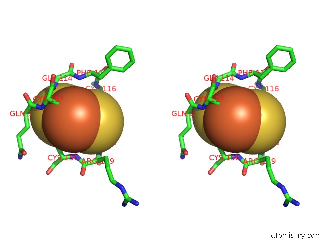
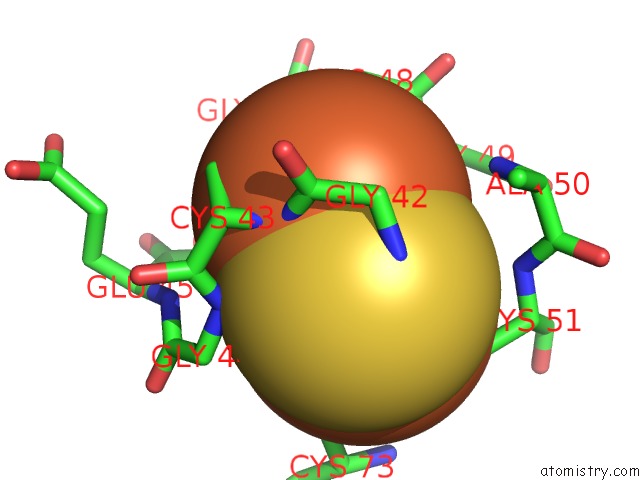
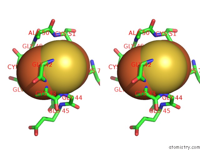
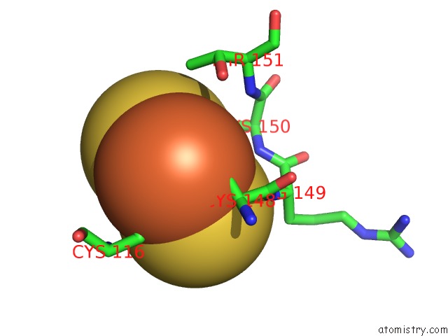
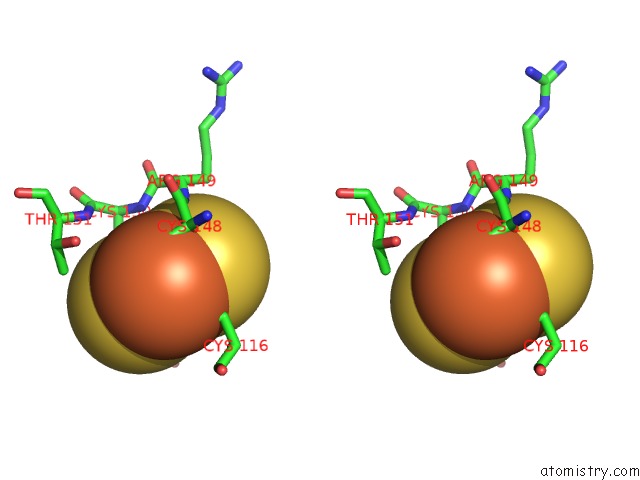
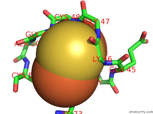
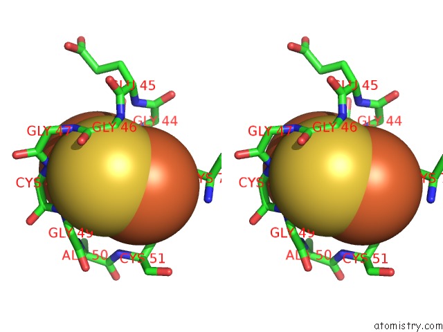
Iron binding site 2 out
of 8 in 3ns1
Go back to
Iron Binding Sites List in 3ns1
Iron binding site 2 out
of 8 in the Crystal Structure of Bovine Xanthine Oxidase in Complex with 6- Mercaptopurine
 Mono view
 Stereo pair view
|
A full contact list of Iron with other atoms in the Fe binding
site number 2 of Crystal Structure of Bovine Xanthine Oxidase in Complex with 6- Mercaptopurine within 5.0Å range:
probe
|
atom
|
residue
|
distance (Å)
|
B
|
Occ
|
A:Fe601
b:14.1
occ:1.00
|
FE2
|
A:FES601
|
0.0
|
14.1
|
1.0
|
S2
|
A:FES601
|
2.2
|
12.6
|
1.0
|
S1
|
A:FES601
|
2.2
|
12.6
|
1.0
|
SG
|
A:CYS116
|
2.2
|
15.9
|
1.0
|
SG
|
A:CYS148
|
2.4
|
15.6
|
1.0
|
FE1
|
A:FES601
|
2.9
|
17.1
|
1.0
|
CB
|
A:CYS148
|
3.3
|
17.2
|
1.0
|
CB
|
A:CYS116
|
3.4
|
15.4
|
1.0
|
CA
|
A:CYS148
|
3.8
|
17.6
|
1.0
|
N
|
A:ARG149
|
4.2
|
19.0
|
1.0
|
N
|
A:CYS116
|
4.3
|
15.6
|
1.0
|
C
|
A:CYS148
|
4.4
|
18.2
|
1.0
|
CA
|
A:CYS116
|
4.4
|
15.3
|
1.0
|
N
|
A:CYS150
|
4.5
|
19.7
|
1.0
|
CB
|
A:CYS150
|
4.6
|
20.5
|
1.0
|
O
|
A:HOH342
|
4.6
|
12.3
|
1.0
|
SG
|
A:CYS150
|
4.8
|
17.6
|
1.0
|
SG
|
A:CYS113
|
4.9
|
15.8
|
1.0
|
C
|
A:CYS116
|
4.9
|
15.2
|
1.0
|
|
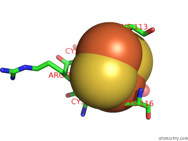
Iron binding site 3 out
of 8 in 3ns1
Go back to
Iron Binding Sites List in 3ns1
Iron binding site 3 out
of 8 in the Crystal Structure of Bovine Xanthine Oxidase in Complex with 6- Mercaptopurine
 Mono view
 Stereo pair view
|
A full contact list of Iron with other atoms in the Fe binding
site number 3 of Crystal Structure of Bovine Xanthine Oxidase in Complex with 6- Mercaptopurine within 5.0Å range:
probe
|
atom
|
residue
|
distance (Å)
|
B
|
Occ
|
A:Fe602
b:17.3
occ:1.00
|
FE1
|
A:FES602
|
0.0
|
17.3
|
1.0
|
S2
|
A:FES602
|
2.2
|
15.5
|
1.0
|
S1
|
A:FES602
|
2.2
|
16.2
|
1.0
|
SG
|
A:CYS51
|
2.2
|
16.2
|
1.0
|
SG
|
A:CYS73
|
2.3
|
17.1
|
1.0
|
FE2
|
A:FES602
|
3.1
|
18.6
|
1.0
|
CB
|
A:CYS73
|
3.2
|
18.9
|
1.0
|
CB
|
A:CYS51
|
3.3
|
16.3
|
1.0
|
N
|
A:CYS73
|
4.1
|
19.5
|
1.0
|
CA
|
A:CYS73
|
4.2
|
19.6
|
1.0
|
CB
|
A:ASN71
|
4.3
|
20.0
|
1.0
|
N
|
A:CYS51
|
4.4
|
16.9
|
1.0
|
OD1
|
A:ASN71
|
4.4
|
22.7
|
1.0
|
CA
|
A:CYS51
|
4.4
|
16.9
|
1.0
|
CG
|
A:ASN71
|
4.5
|
19.8
|
1.0
|
N
|
A:GLY46
|
4.6
|
20.3
|
1.0
|
N
|
A:GLY44
|
4.6
|
18.8
|
1.0
|
CA
|
A:GLY46
|
4.7
|
20.2
|
1.0
|
SG
|
A:CYS43
|
4.7
|
19.5
|
1.0
|
SG
|
A:CYS48
|
4.8
|
15.7
|
1.0
|
CA
|
A:GLY44
|
4.9
|
18.7
|
1.0
|
CA
|
A:ASN71
|
5.0
|
20.4
|
1.0
|
|
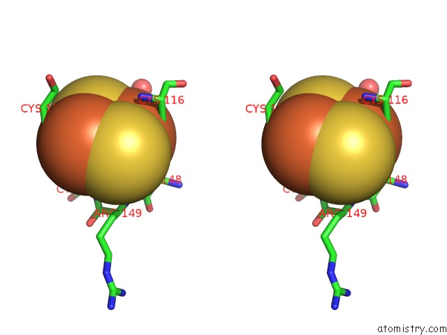
Iron binding site 4 out
of 8 in 3ns1
Go back to
Iron Binding Sites List in 3ns1
Iron binding site 4 out
of 8 in the Crystal Structure of Bovine Xanthine Oxidase in Complex with 6- Mercaptopurine
 Mono view
 Stereo pair view
|
A full contact list of Iron with other atoms in the Fe binding
site number 4 of Crystal Structure of Bovine Xanthine Oxidase in Complex with 6- Mercaptopurine within 5.0Å range:
probe
|
atom
|
residue
|
distance (Å)
|
B
|
Occ
|
A:Fe602
b:18.6
occ:1.00
|
FE2
|
A:FES602
|
0.0
|
18.6
|
1.0
|
SG
|
A:CYS48
|
2.2
|
15.7
|
1.0
|
S2
|
A:FES602
|
2.2
|
15.5
|
1.0
|
S1
|
A:FES602
|
2.2
|
16.2
|
1.0
|
SG
|
A:CYS43
|
2.5
|
19.5
|
1.0
|
FE1
|
A:FES602
|
3.1
|
17.3
|
1.0
|
N
|
A:CYS43
|
3.2
|
17.1
|
1.0
|
CB
|
A:CYS48
|
3.3
|
16.9
|
1.0
|
CB
|
A:CYS43
|
3.5
|
18.1
|
1.0
|
N
|
A:CYS48
|
3.5
|
18.7
|
1.0
|
CA
|
A:CYS48
|
3.8
|
17.2
|
1.0
|
CA
|
A:CYS43
|
3.8
|
18.1
|
1.0
|
N
|
A:GLY44
|
3.9
|
18.8
|
1.0
|
N
|
A:GLY49
|
3.9
|
16.7
|
1.0
|
C
|
A:GLY42
|
4.0
|
17.0
|
1.0
|
CA
|
A:GLY42
|
4.2
|
16.0
|
1.0
|
C
|
A:CYS48
|
4.3
|
16.5
|
1.0
|
N
|
A:GLY42
|
4.3
|
16.0
|
1.0
|
C
|
A:CYS43
|
4.3
|
18.6
|
1.0
|
N
|
A:GLY47
|
4.5
|
20.9
|
1.0
|
N
|
A:ALA50
|
4.5
|
16.2
|
1.0
|
N
|
A:GLY46
|
4.6
|
20.3
|
1.0
|
C
|
A:GLY47
|
4.7
|
20.2
|
1.0
|
SG
|
A:CYS73
|
4.7
|
17.1
|
1.0
|
N
|
A:GLU45
|
4.9
|
20.6
|
1.0
|
SG
|
A:CYS51
|
4.9
|
16.2
|
1.0
|
O
|
A:GLY42
|
4.9
|
16.8
|
1.0
|
CA
|
A:GLY49
|
4.9
|
16.2
|
1.0
|
CB
|
A:ALA50
|
4.9
|
15.0
|
1.0
|
C
|
A:GLY46
|
5.0
|
21.2
|
1.0
|
CA
|
A:GLY46
|
5.0
|
20.2
|
1.0
|
|
Iron binding site 5 out
of 8 in 3ns1
Go back to
Iron Binding Sites List in 3ns1
Iron binding site 5 out
of 8 in the Crystal Structure of Bovine Xanthine Oxidase in Complex with 6- Mercaptopurine
 Mono view
 Stereo pair view
|
A full contact list of Iron with other atoms in the Fe binding
site number 5 of Crystal Structure of Bovine Xanthine Oxidase in Complex with 6- Mercaptopurine within 5.0Å range:
probe
|
atom
|
residue
|
distance (Å)
|
B
|
Occ
|
J:Fe601
b:14.3
occ:1.00
|
FE1
|
J:FES601
|
0.0
|
14.3
|
1.0
|
S1
|
J:FES601
|
2.2
|
15.5
|
1.0
|
S2
|
J:FES601
|
2.2
|
14.7
|
1.0
|
SG
|
J:CYS113
|
2.3
|
17.3
|
1.0
|
SG
|
J:CYS150
|
2.4
|
19.0
|
1.0
|
FE2
|
J:FES601
|
2.9
|
19.5
|
1.0
|
CB
|
J:CYS150
|
3.3
|
20.4
|
1.0
|
CB
|
J:CYS113
|
3.3
|
16.1
|
1.0
|
N
|
J:CYS113
|
3.6
|
16.6
|
1.0
|
CA
|
J:CYS113
|
3.9
|
16.1
|
1.0
|
N
|
J:GLY114
|
3.9
|
15.1
|
1.0
|
N
|
J:CYS150
|
4.0
|
19.6
|
1.0
|
C
|
J:CYS113
|
4.2
|
15.8
|
1.0
|
CA
|
J:CYS150
|
4.3
|
19.6
|
1.0
|
N
|
J:PHE115
|
4.6
|
15.5
|
1.0
|
SG
|
J:CYS148
|
4.6
|
17.2
|
1.0
|
C
|
J:GLN112
|
4.8
|
16.7
|
1.0
|
N
|
J:ARG149
|
4.8
|
19.0
|
1.0
|
SG
|
J:CYS116
|
4.9
|
16.8
|
1.0
|
CB
|
J:GLN112
|
4.9
|
16.0
|
1.0
|
C
|
J:ARG149
|
4.9
|
19.6
|
1.0
|
CA
|
J:GLY114
|
5.0
|
14.9
|
1.0
|
|
Iron binding site 6 out
of 8 in 3ns1
Go back to
Iron Binding Sites List in 3ns1
Iron binding site 6 out
of 8 in the Crystal Structure of Bovine Xanthine Oxidase in Complex with 6- Mercaptopurine
 Mono view
 Stereo pair view
|
A full contact list of Iron with other atoms in the Fe binding
site number 6 of Crystal Structure of Bovine Xanthine Oxidase in Complex with 6- Mercaptopurine within 5.0Å range:
probe
|
atom
|
residue
|
distance (Å)
|
B
|
Occ
|
J:Fe601
b:19.5
occ:1.00
|
FE2
|
J:FES601
|
0.0
|
19.5
|
1.0
|
S2
|
J:FES601
|
2.2
|
14.7
|
1.0
|
S1
|
J:FES601
|
2.2
|
15.5
|
1.0
|
SG
|
J:CYS116
|
2.4
|
16.8
|
1.0
|
SG
|
J:CYS148
|
2.5
|
17.2
|
1.0
|
FE1
|
J:FES601
|
2.9
|
14.3
|
1.0
|
CB
|
J:CYS148
|
3.3
|
17.9
|
1.0
|
CB
|
J:CYS116
|
3.6
|
15.3
|
1.0
|
CA
|
J:CYS148
|
3.7
|
18.0
|
1.0
|
N
|
J:ARG149
|
4.1
|
19.0
|
1.0
|
C
|
J:CYS148
|
4.3
|
18.6
|
1.0
|
N
|
J:CYS150
|
4.3
|
19.6
|
1.0
|
CB
|
J:CYS150
|
4.4
|
20.4
|
1.0
|
N
|
J:CYS116
|
4.5
|
16.0
|
1.0
|
SG
|
J:CYS150
|
4.6
|
19.0
|
1.0
|
CA
|
J:CYS116
|
4.6
|
15.4
|
1.0
|
SG
|
J:CYS113
|
4.8
|
17.3
|
1.0
|
CG2
|
J:THR151
|
4.9
|
18.2
|
1.0
|
CA
|
J:CYS150
|
5.0
|
19.6
|
1.0
|
|
Iron binding site 7 out
of 8 in 3ns1
Go back to
Iron Binding Sites List in 3ns1
Iron binding site 7 out
of 8 in the Crystal Structure of Bovine Xanthine Oxidase in Complex with 6- Mercaptopurine
 Mono view
 Stereo pair view
|
A full contact list of Iron with other atoms in the Fe binding
site number 7 of Crystal Structure of Bovine Xanthine Oxidase in Complex with 6- Mercaptopurine within 5.0Å range:
probe
|
atom
|
residue
|
distance (Å)
|
B
|
Occ
|
J:Fe602
b:19.8
occ:1.00
|
FE1
|
J:FES602
|
0.0
|
19.8
|
1.0
|
S2
|
J:FES602
|
2.2
|
20.5
|
1.0
|
S1
|
J:FES602
|
2.2
|
21.0
|
1.0
|
SG
|
J:CYS51
|
2.2
|
17.2
|
1.0
|
SG
|
J:CYS73
|
2.3
|
18.9
|
1.0
|
FE2
|
J:FES602
|
3.0
|
18.3
|
1.0
|
CB
|
J:CYS73
|
3.2
|
19.0
|
1.0
|
CB
|
J:CYS51
|
3.4
|
16.6
|
1.0
|
N
|
J:CYS73
|
4.2
|
19.3
|
1.0
|
CA
|
J:CYS73
|
4.3
|
19.6
|
1.0
|
CB
|
J:ASN71
|
4.4
|
19.9
|
1.0
|
N
|
J:CYS51
|
4.4
|
16.8
|
1.0
|
CA
|
J:CYS51
|
4.5
|
17.1
|
1.0
|
OD1
|
J:ASN71
|
4.5
|
22.2
|
1.0
|
N
|
J:GLY46
|
4.6
|
20.9
|
1.0
|
CG
|
J:ASN71
|
4.6
|
19.8
|
1.0
|
N
|
J:GLY44
|
4.6
|
18.6
|
1.0
|
SG
|
J:CYS43
|
4.6
|
20.4
|
1.0
|
SG
|
J:CYS48
|
4.7
|
16.2
|
1.0
|
CA
|
J:GLY46
|
4.7
|
20.6
|
1.0
|
N
|
J:GLY49
|
4.9
|
16.1
|
1.0
|
CA
|
J:GLY44
|
4.9
|
18.8
|
1.0
|
|
Iron binding site 8 out
of 8 in 3ns1
Go back to
Iron Binding Sites List in 3ns1
Iron binding site 8 out
of 8 in the Crystal Structure of Bovine Xanthine Oxidase in Complex with 6- Mercaptopurine
 Mono view
 Stereo pair view
|
A full contact list of Iron with other atoms in the Fe binding
site number 8 of Crystal Structure of Bovine Xanthine Oxidase in Complex with 6- Mercaptopurine within 5.0Å range:
probe
|
atom
|
residue
|
distance (Å)
|
B
|
Occ
|
J:Fe602
b:18.3
occ:1.00
|
FE2
|
J:FES602
|
0.0
|
18.3
|
1.0
|
S2
|
J:FES602
|
2.2
|
20.5
|
1.0
|
S1
|
J:FES602
|
2.2
|
21.0
|
1.0
|
SG
|
J:CYS48
|
2.2
|
16.2
|
1.0
|
SG
|
J:CYS43
|
2.4
|
20.4
|
1.0
|
FE1
|
J:FES602
|
3.0
|
19.8
|
1.0
|
N
|
J:CYS43
|
3.2
|
17.0
|
1.0
|
CB
|
J:CYS48
|
3.3
|
16.9
|
1.0
|
CB
|
J:CYS43
|
3.4
|
18.1
|
1.0
|
N
|
J:CYS48
|
3.5
|
18.8
|
1.0
|
CA
|
J:CYS43
|
3.7
|
18.1
|
1.0
|
N
|
J:GLY44
|
3.8
|
18.6
|
1.0
|
CA
|
J:CYS48
|
3.9
|
17.0
|
1.0
|
N
|
J:GLY49
|
3.9
|
16.1
|
1.0
|
C
|
J:GLY42
|
4.0
|
17.1
|
1.0
|
C
|
J:CYS48
|
4.3
|
16.6
|
1.0
|
CA
|
J:GLY42
|
4.3
|
15.7
|
1.0
|
C
|
J:CYS43
|
4.3
|
18.5
|
1.0
|
N
|
J:GLY42
|
4.3
|
15.6
|
1.0
|
N
|
J:GLY47
|
4.4
|
21.2
|
1.0
|
N
|
J:GLY46
|
4.6
|
20.9
|
1.0
|
SG
|
J:CYS73
|
4.6
|
18.9
|
1.0
|
N
|
J:ALA50
|
4.6
|
16.0
|
1.0
|
C
|
J:GLY47
|
4.7
|
20.5
|
1.0
|
N
|
J:GLU45
|
4.8
|
20.5
|
1.0
|
SG
|
J:CYS51
|
4.9
|
17.2
|
1.0
|
CA
|
J:GLY49
|
4.9
|
16.1
|
1.0
|
C
|
J:GLY46
|
4.9
|
21.2
|
1.0
|
CA
|
J:GLY44
|
4.9
|
18.8
|
1.0
|
O
|
J:GLY42
|
4.9
|
16.8
|
1.0
|
CA
|
J:GLY46
|
5.0
|
20.6
|
1.0
|
|
Reference:
H.Cao,
J.M.Pauff,
R.Hille.
Substrate Orientation and Catalytic Specificity in the Action of Xanthine Oxidase: the Sequential Hydroxylation of Hypoxanthine to Uric Acid. J.Biol.Chem. V. 285 28044 2010.
ISSN: ISSN 0021-9258
PubMed: 20615869
DOI: 10.1074/JBC.M110.128561
Page generated: Sun Aug 4 16:40:55 2024
|