Iron »
PDB 3nmk-3nwv »
3nu1 »
Iron in PDB 3nu1: Structure of Holo Form of A Periplasmic Heme Binding Protein
Protein crystallography data
The structure of Structure of Holo Form of A Periplasmic Heme Binding Protein, PDB code: 3nu1
was solved by
D.Mattle,
B.A.Goetz,
J.S.Woo,
K.P.Locher,
with X-Ray Crystallography technique. A brief refinement statistics is given in the table below:
| Resolution Low / High (Å) | 29.64 / 2.50 |
| Space group | P 61 2 2 |
| Cell size a, b, c (Å), α, β, γ (°) | 89.240, 89.240, 288.900, 90.00, 90.00, 120.00 |
| R / Rfree (%) | 19.6 / 23.8 |
Iron Binding Sites:
The binding sites of Iron atom in the Structure of Holo Form of A Periplasmic Heme Binding Protein
(pdb code 3nu1). This binding sites where shown within
5.0 Angstroms radius around Iron atom.
In total 4 binding sites of Iron where determined in the Structure of Holo Form of A Periplasmic Heme Binding Protein, PDB code: 3nu1:
Jump to Iron binding site number: 1; 2; 3; 4;
In total 4 binding sites of Iron where determined in the Structure of Holo Form of A Periplasmic Heme Binding Protein, PDB code: 3nu1:
Jump to Iron binding site number: 1; 2; 3; 4;
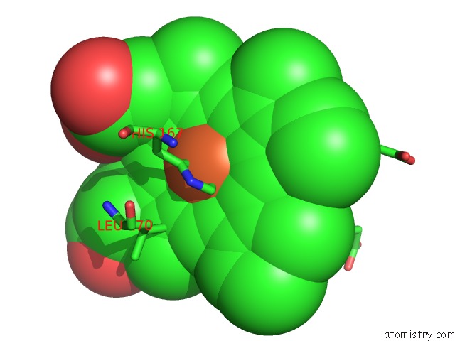
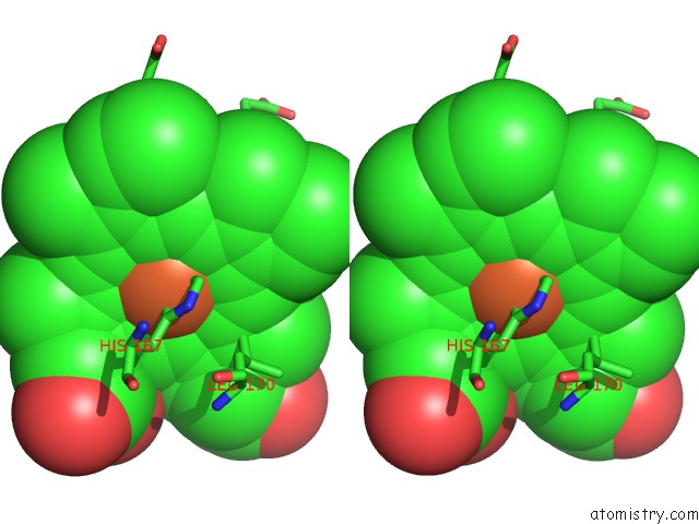
Iron binding site 1 out of 4 in 3nu1
Go back to
Iron binding site 1 out
of 4 in the Structure of Holo Form of A Periplasmic Heme Binding Protein
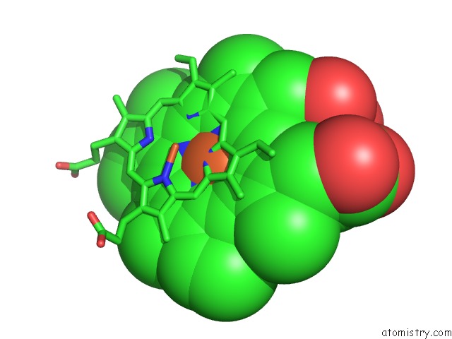
Mono view
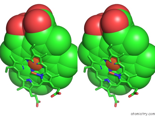
Stereo pair view
Mono view
Stereo pair view
A full contact list of Iron with other atoms in the Fe binding
site number 1 of Structure of Holo Form of A Periplasmic Heme Binding Protein within 5.0Å range:
|
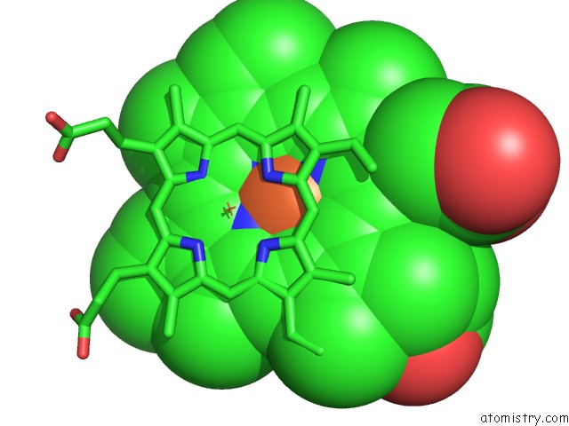
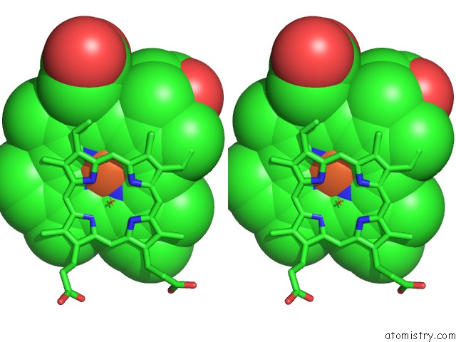
Iron binding site 2 out of 4 in 3nu1
Go back to
Iron binding site 2 out
of 4 in the Structure of Holo Form of A Periplasmic Heme Binding Protein
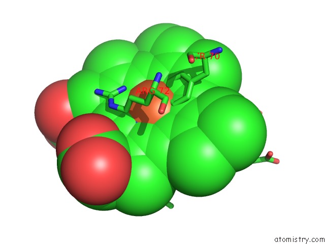
Mono view
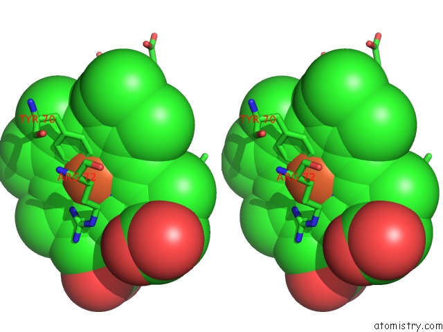
Stereo pair view
Mono view
Stereo pair view
A full contact list of Iron with other atoms in the Fe binding
site number 2 of Structure of Holo Form of A Periplasmic Heme Binding Protein within 5.0Å range:
|
Iron binding site 3 out of 4 in 3nu1
Go back to
Iron binding site 3 out
of 4 in the Structure of Holo Form of A Periplasmic Heme Binding Protein
Mono view
Stereo pair view
Mono view
Stereo pair view
A full contact list of Iron with other atoms in the Fe binding
site number 3 of Structure of Holo Form of A Periplasmic Heme Binding Protein within 5.0Å range:
|
Iron binding site 4 out of 4 in 3nu1
Go back to
Iron binding site 4 out
of 4 in the Structure of Holo Form of A Periplasmic Heme Binding Protein
Mono view
Stereo pair view
Mono view
Stereo pair view
A full contact list of Iron with other atoms in the Fe binding
site number 4 of Structure of Holo Form of A Periplasmic Heme Binding Protein within 5.0Å range:
|
Reference:
D.Mattle,
A.Zeltina,
J.S.Woo,
B.A.Goetz,
K.P.Locher.
Two Stacked Heme Molecules in the Binding Pocket of the Periplasmic Heme-Binding Protein Hmut From Yersinia Pestis. J.Mol.Biol. V. 404 220 2010.
ISSN: ISSN 0022-2836
PubMed: 20888343
DOI: 10.1016/J.JMB.2010.09.005
Page generated: Tue Aug 5 04:56:37 2025
ISSN: ISSN 0022-2836
PubMed: 20888343
DOI: 10.1016/J.JMB.2010.09.005
Last articles
Fe in 3VM1Fe in 3VM0
Fe in 3VM9
Fe in 3VM4
Fe in 3VLY
Fe in 3VLX
Fe in 3VLZ
Fe in 3VKT
Fe in 3VLL
Fe in 3VLM