Iron »
PDB 3nmk-3nwv »
3nv6 »
Iron in PDB 3nv6: Crystal Structure of Camphor-Bound CYP101D2
Protein crystallography data
The structure of Crystal Structure of Camphor-Bound CYP101D2, PDB code: 3nv6
was solved by
W.Yang,
S.G.Bell,
H.Wang,
W.H.Zhou,
M.Bartlam,
L.L.Wong,
Z.Rao,
with X-Ray Crystallography technique. A brief refinement statistics is given in the table below:
| Resolution Low / High (Å) | 47.64 / 2.20 |
| Space group | P 32 2 1 |
| Cell size a, b, c (Å), α, β, γ (°) | 86.080, 86.080, 123.867, 90.00, 90.00, 120.00 |
| R / Rfree (%) | 18.9 / 25.5 |
Iron Binding Sites:
The binding sites of Iron atom in the Crystal Structure of Camphor-Bound CYP101D2
(pdb code 3nv6). This binding sites where shown within
5.0 Angstroms radius around Iron atom.
In total only one binding site of Iron was determined in the Crystal Structure of Camphor-Bound CYP101D2, PDB code: 3nv6:
In total only one binding site of Iron was determined in the Crystal Structure of Camphor-Bound CYP101D2, PDB code: 3nv6:
Iron binding site 1 out of 1 in 3nv6
Go back to
Iron binding site 1 out
of 1 in the Crystal Structure of Camphor-Bound CYP101D2
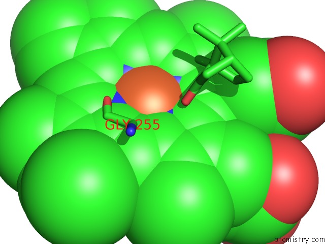
Mono view
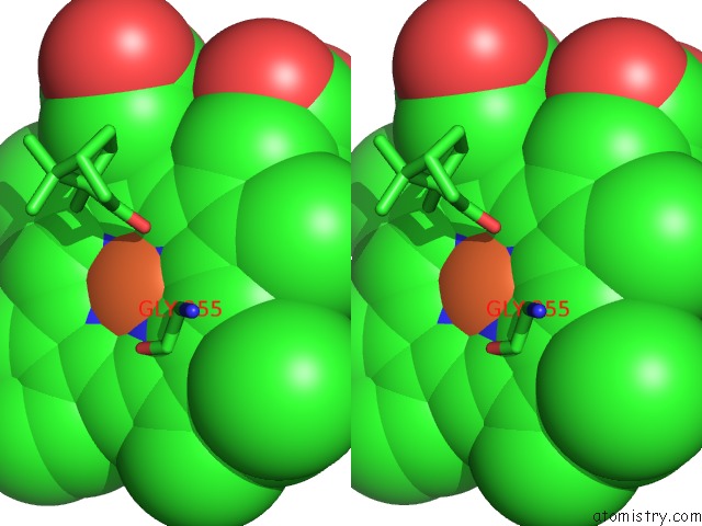
Stereo pair view
Mono view
Stereo pair view
A full contact list of Iron with other atoms in the Fe binding
site number 1 of Crystal Structure of Camphor-Bound CYP101D2 within 5.0Å range:
|
Reference:
W.Yang,
S.G.Bell,
H.Wang,
W.Zhou,
M.Bartlam,
L.L.Wong,
Z.Rao.
The Structure of CYP101D2 Unveils A Potential Path For Substrate Entry Into the Active Site Biochem.J. V. 433 85 2011.
ISSN: ISSN 0264-6021
PubMed: 20950270
DOI: 10.1042/BJ20101017
Page generated: Tue Aug 5 04:57:35 2025
ISSN: ISSN 0264-6021
PubMed: 20950270
DOI: 10.1042/BJ20101017
Last articles
Fe in 3VM0Fe in 3VM9
Fe in 3VM4
Fe in 3VLY
Fe in 3VLX
Fe in 3VLZ
Fe in 3VKT
Fe in 3VLL
Fe in 3VLM
Fe in 3VLK