Iron »
PDB 3nmk-3nwv »
3nvr »
Iron in PDB 3nvr: Modulating Heme Redox Potential Through Protein-Induced Porphyrin Distortion
Protein crystallography data
The structure of Modulating Heme Redox Potential Through Protein-Induced Porphyrin Distortion, PDB code: 3nvr
was solved by
C.Olea Jr.,
J.Kuriyan,
M.A.Marletta,
with X-Ray Crystallography technique. A brief refinement statistics is given in the table below:
| Resolution Low / High (Å) | 49.48 / 2.15 |
| Space group | P 21 21 2 |
| Cell size a, b, c (Å), α, β, γ (°) | 79.825, 126.095, 42.742, 90.00, 90.00, 90.00 |
| R / Rfree (%) | 21.7 / 26 |
Other elements in 3nvr:
The structure of Modulating Heme Redox Potential Through Protein-Induced Porphyrin Distortion also contains other interesting chemical elements:
| Chlorine | (Cl) | 2 atoms |
Iron Binding Sites:
The binding sites of Iron atom in the Modulating Heme Redox Potential Through Protein-Induced Porphyrin Distortion
(pdb code 3nvr). This binding sites where shown within
5.0 Angstroms radius around Iron atom.
In total 2 binding sites of Iron where determined in the Modulating Heme Redox Potential Through Protein-Induced Porphyrin Distortion, PDB code: 3nvr:
Jump to Iron binding site number: 1; 2;
In total 2 binding sites of Iron where determined in the Modulating Heme Redox Potential Through Protein-Induced Porphyrin Distortion, PDB code: 3nvr:
Jump to Iron binding site number: 1; 2;
Iron binding site 1 out of 2 in 3nvr
Go back to
Iron binding site 1 out
of 2 in the Modulating Heme Redox Potential Through Protein-Induced Porphyrin Distortion
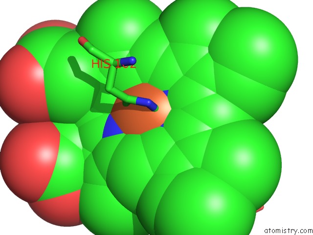
Mono view
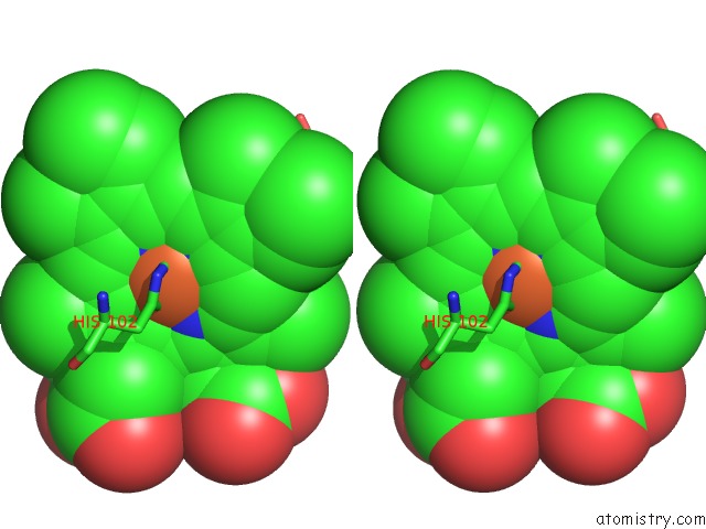
Stereo pair view
Mono view
Stereo pair view
A full contact list of Iron with other atoms in the Fe binding
site number 1 of Modulating Heme Redox Potential Through Protein-Induced Porphyrin Distortion within 5.0Å range:
|
Iron binding site 2 out of 2 in 3nvr
Go back to
Iron binding site 2 out
of 2 in the Modulating Heme Redox Potential Through Protein-Induced Porphyrin Distortion
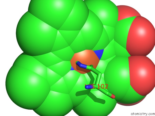
Mono view
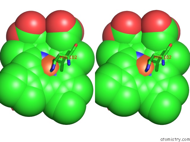
Stereo pair view
Mono view
Stereo pair view
A full contact list of Iron with other atoms in the Fe binding
site number 2 of Modulating Heme Redox Potential Through Protein-Induced Porphyrin Distortion within 5.0Å range:
|
Reference:
C.Olea,
J.Kuriyan,
M.A.Marletta.
Modulating Heme Redox Potential Through Protein-Induced Porphyrin Distortion J.Am.Chem.Soc. V. 132 12794 2010.
ISSN: ISSN 0002-7863
PubMed: 20735135
DOI: 10.1021/JA106252B
Page generated: Tue Aug 5 04:57:56 2025
ISSN: ISSN 0002-7863
PubMed: 20735135
DOI: 10.1021/JA106252B
Last articles
Fe in 3VM1Fe in 3VM0
Fe in 3VM9
Fe in 3VM4
Fe in 3VLY
Fe in 3VLX
Fe in 3VLZ
Fe in 3VKT
Fe in 3VLL
Fe in 3VLM