Iron »
PDB 3nmk-3nwv »
3nvz »
Iron in PDB 3nvz: Crystal Structure of Bovine Xanthine Oxidase in Complex with Indole-3- Aldehyde
Enzymatic activity of Crystal Structure of Bovine Xanthine Oxidase in Complex with Indole-3- Aldehyde
All present enzymatic activity of Crystal Structure of Bovine Xanthine Oxidase in Complex with Indole-3- Aldehyde:
1.17.1.4; 1.17.3.2;
1.17.1.4; 1.17.3.2;
Protein crystallography data
The structure of Crystal Structure of Bovine Xanthine Oxidase in Complex with Indole-3- Aldehyde, PDB code: 3nvz
was solved by
H.Cao,
R.Hille,
with X-Ray Crystallography technique. A brief refinement statistics is given in the table below:
| Resolution Low / High (Å) | 45.20 / 1.60 |
| Space group | P 1 21 1 |
| Cell size a, b, c (Å), α, β, γ (°) | 133.409, 73.704, 138.922, 90.00, 97.12, 90.00 |
| R / Rfree (%) | 21.5 / 24.6 |
Other elements in 3nvz:
The structure of Crystal Structure of Bovine Xanthine Oxidase in Complex with Indole-3- Aldehyde also contains other interesting chemical elements:
| Molybdenum | (Mo) | 2 atoms |
Iron Binding Sites:
The binding sites of Iron atom in the Crystal Structure of Bovine Xanthine Oxidase in Complex with Indole-3- Aldehyde
(pdb code 3nvz). This binding sites where shown within
5.0 Angstroms radius around Iron atom.
In total 8 binding sites of Iron where determined in the Crystal Structure of Bovine Xanthine Oxidase in Complex with Indole-3- Aldehyde, PDB code: 3nvz:
Jump to Iron binding site number: 1; 2; 3; 4; 5; 6; 7; 8;
In total 8 binding sites of Iron where determined in the Crystal Structure of Bovine Xanthine Oxidase in Complex with Indole-3- Aldehyde, PDB code: 3nvz:
Jump to Iron binding site number: 1; 2; 3; 4; 5; 6; 7; 8;
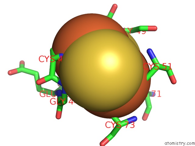
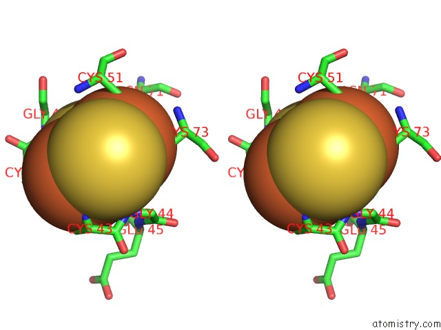
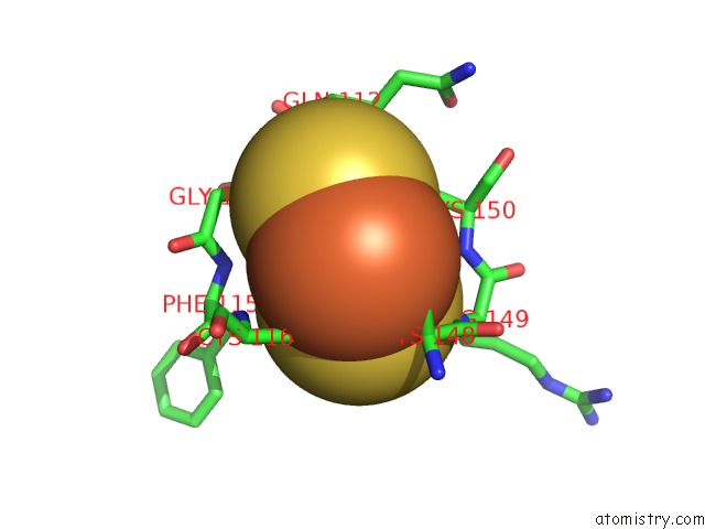
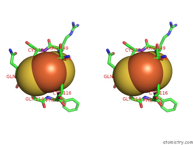
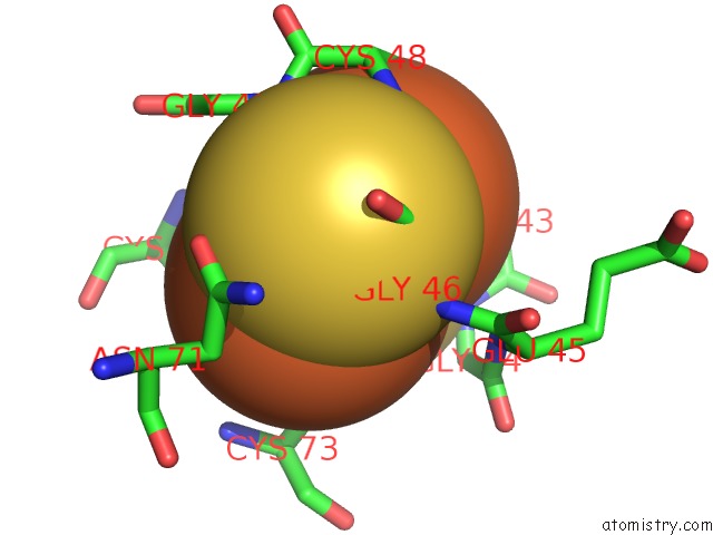
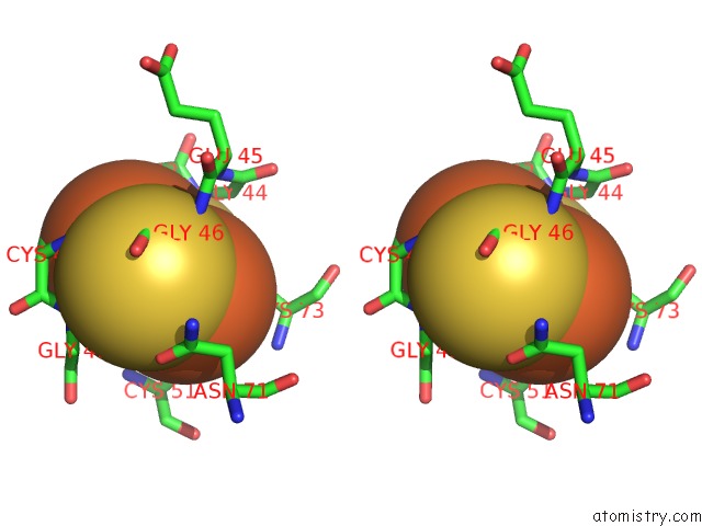
Iron binding site 1 out of 8 in 3nvz
Go back to
Iron binding site 1 out
of 8 in the Crystal Structure of Bovine Xanthine Oxidase in Complex with Indole-3- Aldehyde
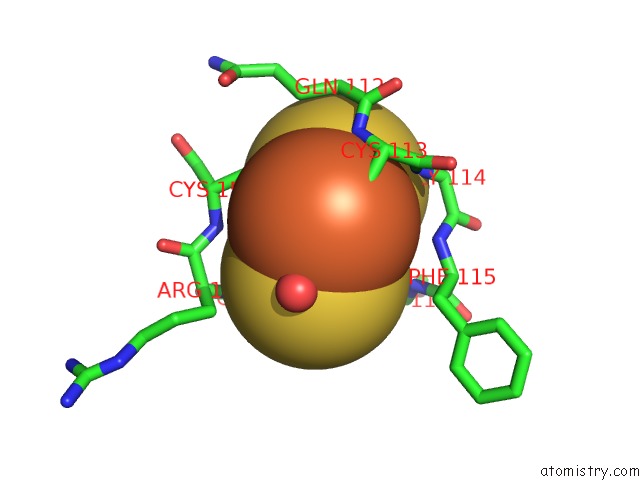
Mono view
Stereo pair view
Mono view
Stereo pair view
A full contact list of Iron with other atoms in the Fe binding
site number 1 of Crystal Structure of Bovine Xanthine Oxidase in Complex with Indole-3- Aldehyde within 5.0Å range:
|
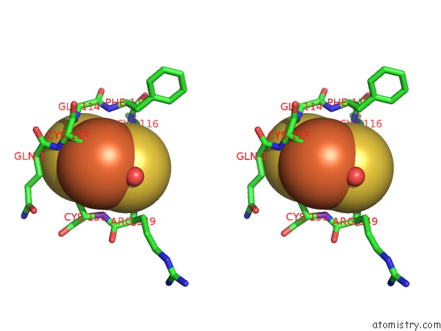
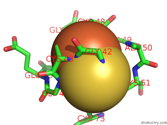
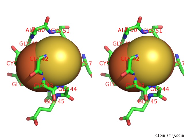
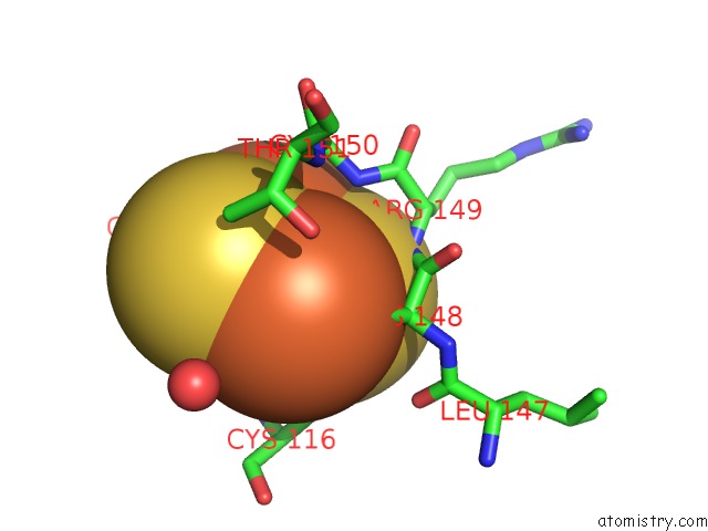
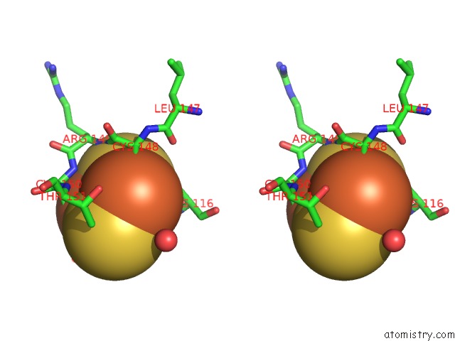
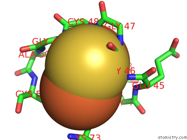
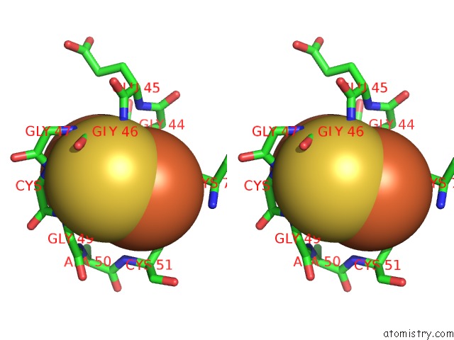
Iron binding site 2 out of 8 in 3nvz
Go back to
Iron binding site 2 out
of 8 in the Crystal Structure of Bovine Xanthine Oxidase in Complex with Indole-3- Aldehyde
Mono view
Stereo pair view
Mono view
Stereo pair view
A full contact list of Iron with other atoms in the Fe binding
site number 2 of Crystal Structure of Bovine Xanthine Oxidase in Complex with Indole-3- Aldehyde within 5.0Å range:
|
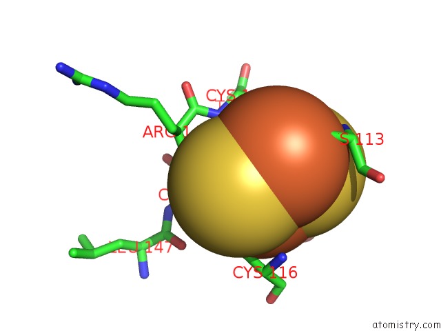
Iron binding site 3 out of 8 in 3nvz
Go back to
Iron binding site 3 out
of 8 in the Crystal Structure of Bovine Xanthine Oxidase in Complex with Indole-3- Aldehyde
Mono view
Stereo pair view
Mono view
Stereo pair view
A full contact list of Iron with other atoms in the Fe binding
site number 3 of Crystal Structure of Bovine Xanthine Oxidase in Complex with Indole-3- Aldehyde within 5.0Å range:
|
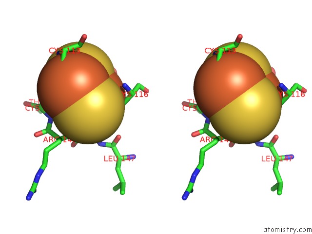
Iron binding site 4 out of 8 in 3nvz
Go back to
Iron binding site 4 out
of 8 in the Crystal Structure of Bovine Xanthine Oxidase in Complex with Indole-3- Aldehyde
Mono view
Stereo pair view
Mono view
Stereo pair view
A full contact list of Iron with other atoms in the Fe binding
site number 4 of Crystal Structure of Bovine Xanthine Oxidase in Complex with Indole-3- Aldehyde within 5.0Å range:
|
Iron binding site 5 out of 8 in 3nvz
Go back to
Iron binding site 5 out
of 8 in the Crystal Structure of Bovine Xanthine Oxidase in Complex with Indole-3- Aldehyde
Mono view
Stereo pair view
Mono view
Stereo pair view
A full contact list of Iron with other atoms in the Fe binding
site number 5 of Crystal Structure of Bovine Xanthine Oxidase in Complex with Indole-3- Aldehyde within 5.0Å range:
|
Iron binding site 6 out of 8 in 3nvz
Go back to
Iron binding site 6 out
of 8 in the Crystal Structure of Bovine Xanthine Oxidase in Complex with Indole-3- Aldehyde
Mono view
Stereo pair view
Mono view
Stereo pair view
A full contact list of Iron with other atoms in the Fe binding
site number 6 of Crystal Structure of Bovine Xanthine Oxidase in Complex with Indole-3- Aldehyde within 5.0Å range:
|
Iron binding site 7 out of 8 in 3nvz
Go back to
Iron binding site 7 out
of 8 in the Crystal Structure of Bovine Xanthine Oxidase in Complex with Indole-3- Aldehyde
Mono view
Stereo pair view
Mono view
Stereo pair view
A full contact list of Iron with other atoms in the Fe binding
site number 7 of Crystal Structure of Bovine Xanthine Oxidase in Complex with Indole-3- Aldehyde within 5.0Å range:
|
Iron binding site 8 out of 8 in 3nvz
Go back to
Iron binding site 8 out
of 8 in the Crystal Structure of Bovine Xanthine Oxidase in Complex with Indole-3- Aldehyde
Mono view
Stereo pair view
Mono view
Stereo pair view
A full contact list of Iron with other atoms in the Fe binding
site number 8 of Crystal Structure of Bovine Xanthine Oxidase in Complex with Indole-3- Aldehyde within 5.0Å range:
|
Reference:
H.Cao,
J.Hall,
R.Hille.
Substrate Orientation and Specificity in Xanthine Oxidase: Crystal Structures of the Enzyme in Complex with Indole-3-Acetaldehyde and Guanine. Biochemistry V. 53 533 2014.
ISSN: ISSN 0006-2960
PubMed: 24397336
DOI: 10.1021/BI401465U
Page generated: Tue Aug 5 05:00:26 2025
ISSN: ISSN 0006-2960
PubMed: 24397336
DOI: 10.1021/BI401465U
Last articles
Fe in 3VYTFe in 3VYU
Fe in 3W1W
Fe in 3W08
Fe in 3VYS
Fe in 3VYR
Fe in 3VV9
Fe in 3VVA
Fe in 3VYM
Fe in 3VXJ