Iron »
PDB 3nxu-3ol5 »
3o0r »
Iron in PDB 3o0r: Crystal Structure of Nitric Oxide Reductase From Pseudomonas Aeruginosa in Complex with Antibody Fragment
Enzymatic activity of Crystal Structure of Nitric Oxide Reductase From Pseudomonas Aeruginosa in Complex with Antibody Fragment
All present enzymatic activity of Crystal Structure of Nitric Oxide Reductase From Pseudomonas Aeruginosa in Complex with Antibody Fragment:
1.7.99.7;
1.7.99.7;
Protein crystallography data
The structure of Crystal Structure of Nitric Oxide Reductase From Pseudomonas Aeruginosa in Complex with Antibody Fragment, PDB code: 3o0r
was solved by
T.Hino,
Y.Matsumoto,
S.Nagano,
H.Sugimoto,
Y.Fukumori,
T.Murata,
S.Iwata,
Y.Shiro,
with X-Ray Crystallography technique. A brief refinement statistics is given in the table below:
| Resolution Low / High (Å) | 20.00 / 2.70 |
| Space group | P 21 21 21 |
| Cell size a, b, c (Å), α, β, γ (°) | 90.470, 104.520, 195.360, 90.00, 90.00, 90.00 |
| R / Rfree (%) | 18.5 / 24.7 |
Other elements in 3o0r:
The structure of Crystal Structure of Nitric Oxide Reductase From Pseudomonas Aeruginosa in Complex with Antibody Fragment also contains other interesting chemical elements:
| Calcium | (Ca) | 1 atom |
Iron Binding Sites:
The binding sites of Iron atom in the Crystal Structure of Nitric Oxide Reductase From Pseudomonas Aeruginosa in Complex with Antibody Fragment
(pdb code 3o0r). This binding sites where shown within
5.0 Angstroms radius around Iron atom.
In total 4 binding sites of Iron where determined in the Crystal Structure of Nitric Oxide Reductase From Pseudomonas Aeruginosa in Complex with Antibody Fragment, PDB code: 3o0r:
Jump to Iron binding site number: 1; 2; 3; 4;
In total 4 binding sites of Iron where determined in the Crystal Structure of Nitric Oxide Reductase From Pseudomonas Aeruginosa in Complex with Antibody Fragment, PDB code: 3o0r:
Jump to Iron binding site number: 1; 2; 3; 4;
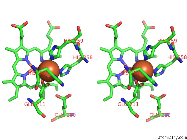
Iron binding site 1 out of 4 in 3o0r
Go back to
Iron binding site 1 out
of 4 in the Crystal Structure of Nitric Oxide Reductase From Pseudomonas Aeruginosa in Complex with Antibody Fragment

Mono view
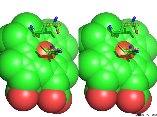
Stereo pair view
Mono view
Stereo pair view
A full contact list of Iron with other atoms in the Fe binding
site number 1 of Crystal Structure of Nitric Oxide Reductase From Pseudomonas Aeruginosa in Complex with Antibody Fragment within 5.0Å range:
|
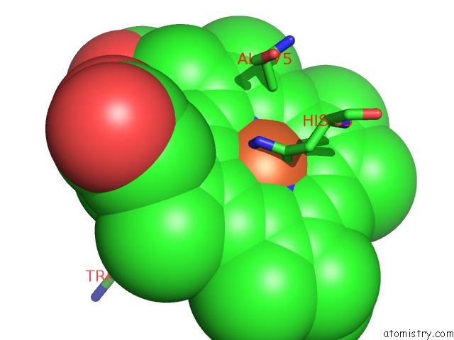
Iron binding site 2 out of 4 in 3o0r
Go back to
Iron binding site 2 out
of 4 in the Crystal Structure of Nitric Oxide Reductase From Pseudomonas Aeruginosa in Complex with Antibody Fragment
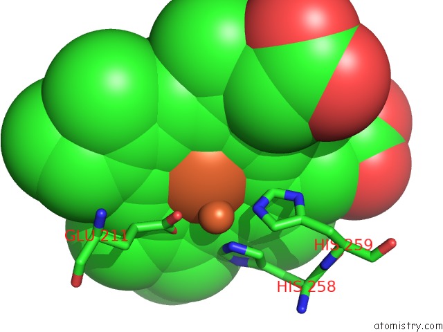
Mono view
Stereo pair view
Mono view
Stereo pair view
A full contact list of Iron with other atoms in the Fe binding
site number 2 of Crystal Structure of Nitric Oxide Reductase From Pseudomonas Aeruginosa in Complex with Antibody Fragment within 5.0Å range:
|
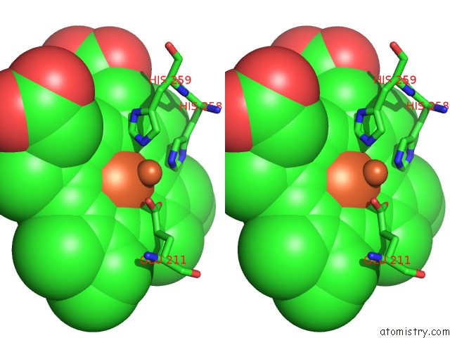
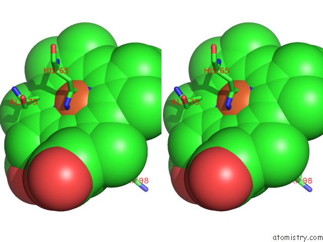
Iron binding site 3 out of 4 in 3o0r
Go back to
Iron binding site 3 out
of 4 in the Crystal Structure of Nitric Oxide Reductase From Pseudomonas Aeruginosa in Complex with Antibody Fragment
Mono view
Stereo pair view
Mono view
Stereo pair view
A full contact list of Iron with other atoms in the Fe binding
site number 3 of Crystal Structure of Nitric Oxide Reductase From Pseudomonas Aeruginosa in Complex with Antibody Fragment within 5.0Å range:
|
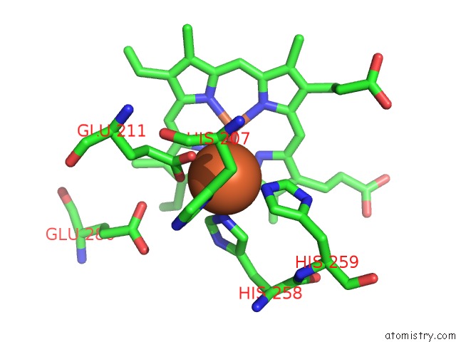
Iron binding site 4 out of 4 in 3o0r
Go back to
Iron binding site 4 out
of 4 in the Crystal Structure of Nitric Oxide Reductase From Pseudomonas Aeruginosa in Complex with Antibody Fragment
Mono view
Stereo pair view
Mono view
Stereo pair view
A full contact list of Iron with other atoms in the Fe binding
site number 4 of Crystal Structure of Nitric Oxide Reductase From Pseudomonas Aeruginosa in Complex with Antibody Fragment within 5.0Å range:
|
Reference:
T.Hino,
Y.Matsumoto,
S.Nagano,
H.Sugimoto,
Y.Fukumori,
T.Murata,
S.Iwata,
Y.Shiro.
Structural Basis of Biological N2O Generation By Bacterial Nitric Oxide Reductase Science V. 330 1666 2010.
ISSN: ISSN 0036-8075
PubMed: 21109633
DOI: 10.1126/SCIENCE.1195591
Page generated: Sun Aug 4 16:56:14 2024
ISSN: ISSN 0036-8075
PubMed: 21109633
DOI: 10.1126/SCIENCE.1195591
Last articles
Zn in 9MJ5Zn in 9HNW
Zn in 9G0L
Zn in 9FNE
Zn in 9DZN
Zn in 9E0I
Zn in 9D32
Zn in 9DAK
Zn in 8ZXC
Zn in 8ZUF