Iron »
PDB 3nxu-3ol5 »
3oft »
Iron in PDB 3oft: Crystal Structure of Cytochrome P450 CYP101C1
Protein crystallography data
The structure of Crystal Structure of Cytochrome P450 CYP101C1, PDB code: 3oft
was solved by
W.Zhou,
M.Ma,
S.G.Bell,
W.Yang,
Y.Hao,
N.H.Rees,
M.Bartlam,
L.-L.Wong,
Z.Rao,
with X-Ray Crystallography technique. A brief refinement statistics is given in the table below:
| Resolution Low / High (Å) | 50.00 / 1.90 |
| Space group | P 1 21 1 |
| Cell size a, b, c (Å), α, β, γ (°) | 57.986, 150.148, 68.454, 90.00, 99.89, 90.00 |
| R / Rfree (%) | 17.4 / 23.2 |
Iron Binding Sites:
The binding sites of Iron atom in the Crystal Structure of Cytochrome P450 CYP101C1
(pdb code 3oft). This binding sites where shown within
5.0 Angstroms radius around Iron atom.
In total 3 binding sites of Iron where determined in the Crystal Structure of Cytochrome P450 CYP101C1, PDB code: 3oft:
Jump to Iron binding site number: 1; 2; 3;
In total 3 binding sites of Iron where determined in the Crystal Structure of Cytochrome P450 CYP101C1, PDB code: 3oft:
Jump to Iron binding site number: 1; 2; 3;
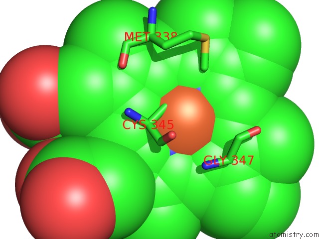
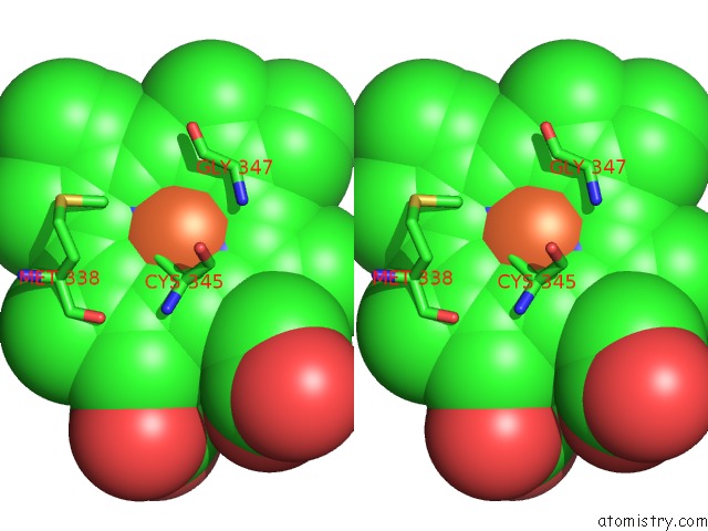
Iron binding site 1 out of 3 in 3oft
Go back to
Iron binding site 1 out
of 3 in the Crystal Structure of Cytochrome P450 CYP101C1
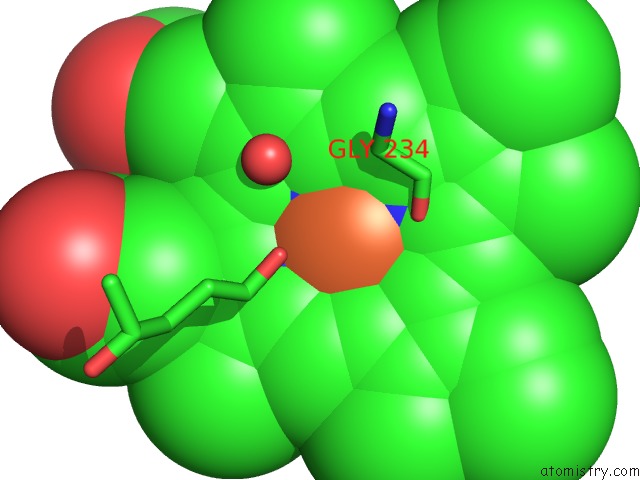
Mono view
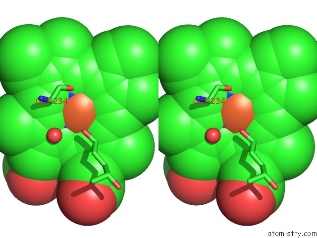
Stereo pair view
Mono view
Stereo pair view
A full contact list of Iron with other atoms in the Fe binding
site number 1 of Crystal Structure of Cytochrome P450 CYP101C1 within 5.0Å range:
|
Iron binding site 2 out of 3 in 3oft
Go back to
Iron binding site 2 out
of 3 in the Crystal Structure of Cytochrome P450 CYP101C1
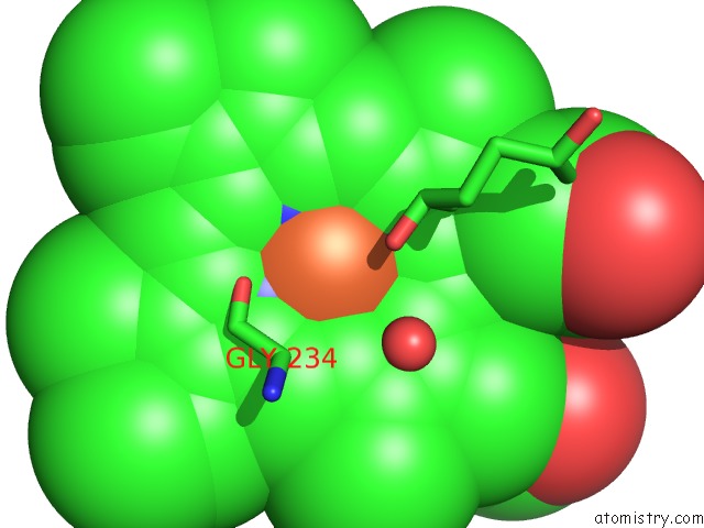
Mono view
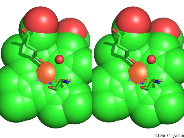
Stereo pair view
Mono view
Stereo pair view
A full contact list of Iron with other atoms in the Fe binding
site number 2 of Crystal Structure of Cytochrome P450 CYP101C1 within 5.0Å range:
|
Iron binding site 3 out of 3 in 3oft
Go back to
Iron binding site 3 out
of 3 in the Crystal Structure of Cytochrome P450 CYP101C1
Mono view
Stereo pair view
Mono view
Stereo pair view
A full contact list of Iron with other atoms in the Fe binding
site number 3 of Crystal Structure of Cytochrome P450 CYP101C1 within 5.0Å range:
|
Reference:
M.Ma,
S.G.Bell,
W.Yang,
Y.Hao,
N.H.Rees,
M.Bartlam,
W.Zhou,
L.L.Wong,
Z.Rao.
Structural Analysis of CYP101C1 From Novosphingobium Aromaticivorans DSM12444. Chembiochem V. 12 88 2011.
ISSN: ISSN 1439-4227
PubMed: 21154803
DOI: 10.1002/CBIC.201000537
Page generated: Sun Aug 4 17:04:23 2024
ISSN: ISSN 1439-4227
PubMed: 21154803
DOI: 10.1002/CBIC.201000537
Last articles
Zn in 9MJ5Zn in 9HNW
Zn in 9G0L
Zn in 9FNE
Zn in 9DZN
Zn in 9E0I
Zn in 9D32
Zn in 9DAK
Zn in 8ZXC
Zn in 8ZUF