Iron »
PDB 3nxu-3ol5 »
3ojt »
Iron in PDB 3ojt: Structure of Native Fe-Containing Homoprotocatechuate 2,3-Dioxygenase at 1.70 Ang Resolution
Protein crystallography data
The structure of Structure of Native Fe-Containing Homoprotocatechuate 2,3-Dioxygenase at 1.70 Ang Resolution, PDB code: 3ojt
was solved by
A.J.Fielding,
E.G.Kovaleva,
E.R.Farquhar,
J.D.Lipscomb,
L.Que Jr.,
with X-Ray Crystallography technique. A brief refinement statistics is given in the table below:
| Resolution Low / High (Å) | 59.49 / 1.70 |
| Space group | P 21 21 2 |
| Cell size a, b, c (Å), α, β, γ (°) | 110.747, 151.229, 96.385, 90.00, 90.00, 90.00 |
| R / Rfree (%) | 16 / 18.8 |
Other elements in 3ojt:
The structure of Structure of Native Fe-Containing Homoprotocatechuate 2,3-Dioxygenase at 1.70 Ang Resolution also contains other interesting chemical elements:
| Chlorine | (Cl) | 4 atoms |
| Calcium | (Ca) | 1 atom |
Iron Binding Sites:
The binding sites of Iron atom in the Structure of Native Fe-Containing Homoprotocatechuate 2,3-Dioxygenase at 1.70 Ang Resolution
(pdb code 3ojt). This binding sites where shown within
5.0 Angstroms radius around Iron atom.
In total 4 binding sites of Iron where determined in the Structure of Native Fe-Containing Homoprotocatechuate 2,3-Dioxygenase at 1.70 Ang Resolution, PDB code: 3ojt:
Jump to Iron binding site number: 1; 2; 3; 4;
In total 4 binding sites of Iron where determined in the Structure of Native Fe-Containing Homoprotocatechuate 2,3-Dioxygenase at 1.70 Ang Resolution, PDB code: 3ojt:
Jump to Iron binding site number: 1; 2; 3; 4;
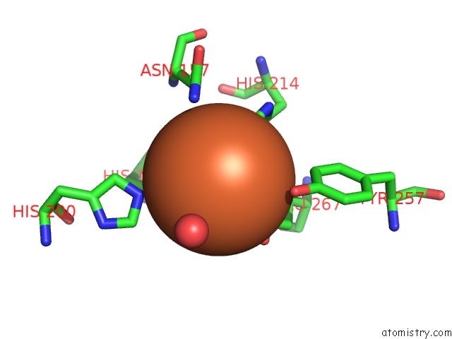
Iron binding site 1 out of 4 in 3ojt
Go back to
Iron binding site 1 out
of 4 in the Structure of Native Fe-Containing Homoprotocatechuate 2,3-Dioxygenase at 1.70 Ang Resolution
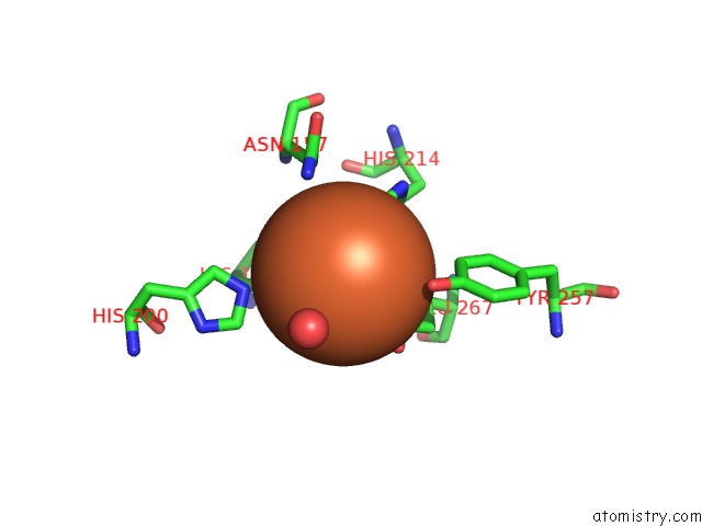
Mono view
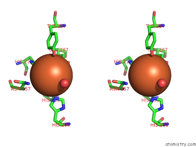
Stereo pair view
Mono view
Stereo pair view
A full contact list of Iron with other atoms in the Fe binding
site number 1 of Structure of Native Fe-Containing Homoprotocatechuate 2,3-Dioxygenase at 1.70 Ang Resolution within 5.0Å range:
|
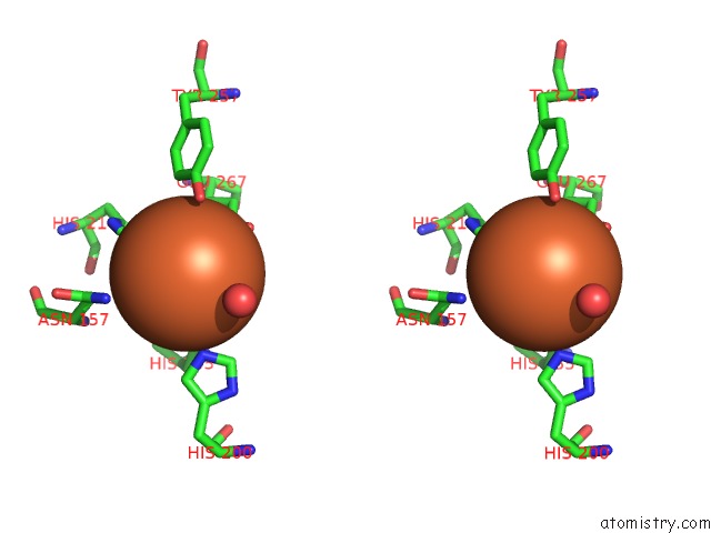
Iron binding site 2 out of 4 in 3ojt
Go back to
Iron binding site 2 out
of 4 in the Structure of Native Fe-Containing Homoprotocatechuate 2,3-Dioxygenase at 1.70 Ang Resolution
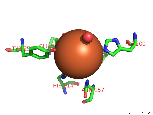
Mono view
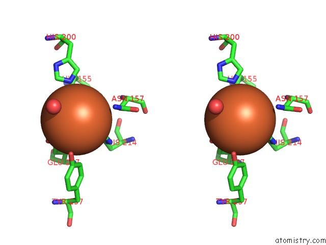
Stereo pair view
Mono view
Stereo pair view
A full contact list of Iron with other atoms in the Fe binding
site number 2 of Structure of Native Fe-Containing Homoprotocatechuate 2,3-Dioxygenase at 1.70 Ang Resolution within 5.0Å range:
|
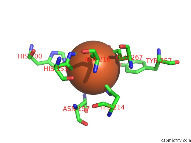
Iron binding site 3 out of 4 in 3ojt
Go back to
Iron binding site 3 out
of 4 in the Structure of Native Fe-Containing Homoprotocatechuate 2,3-Dioxygenase at 1.70 Ang Resolution
Mono view
Stereo pair view
Mono view
Stereo pair view
A full contact list of Iron with other atoms in the Fe binding
site number 3 of Structure of Native Fe-Containing Homoprotocatechuate 2,3-Dioxygenase at 1.70 Ang Resolution within 5.0Å range:
|
Iron binding site 4 out of 4 in 3ojt
Go back to
Iron binding site 4 out
of 4 in the Structure of Native Fe-Containing Homoprotocatechuate 2,3-Dioxygenase at 1.70 Ang Resolution
Mono view
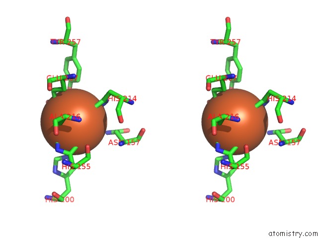
Stereo pair view
Mono view
Stereo pair view
A full contact list of Iron with other atoms in the Fe binding
site number 4 of Structure of Native Fe-Containing Homoprotocatechuate 2,3-Dioxygenase at 1.70 Ang Resolution within 5.0Å range:
|
Reference:
A.J.Fielding,
E.G.Kovaleva,
E.R.Farquhar,
J.D.Lipscomb,
L.Que.
A Hyperactive Cobalt-Substituted Extradiol-Cleaving Catechol Dioxygenase. J.Biol.Inorg.Chem. V. 16 341 2011.
ISSN: ISSN 0949-8257
PubMed: 21153851
DOI: 10.1007/S00775-010-0732-0
Page generated: Sun Aug 4 17:07:49 2024
ISSN: ISSN 0949-8257
PubMed: 21153851
DOI: 10.1007/S00775-010-0732-0
Last articles
Zn in 9MJ5Zn in 9HNW
Zn in 9G0L
Zn in 9FNE
Zn in 9DZN
Zn in 9E0I
Zn in 9D32
Zn in 9DAK
Zn in 8ZXC
Zn in 8ZUF