Iron »
PDB 3om3-3p4q »
3omi »
Iron in PDB 3omi: Catalytic Core Subunits (I and II) of Cytochrome C Oxidase From Rhodobacter Sphaeroides with D132A Mutation
Enzymatic activity of Catalytic Core Subunits (I and II) of Cytochrome C Oxidase From Rhodobacter Sphaeroides with D132A Mutation
All present enzymatic activity of Catalytic Core Subunits (I and II) of Cytochrome C Oxidase From Rhodobacter Sphaeroides with D132A Mutation:
1.9.3.1;
1.9.3.1;
Protein crystallography data
The structure of Catalytic Core Subunits (I and II) of Cytochrome C Oxidase From Rhodobacter Sphaeroides with D132A Mutation, PDB code: 3omi
was solved by
J.Liu,
L.Qin,
S.Ferguson-Miller,
with X-Ray Crystallography technique. A brief refinement statistics is given in the table below:
| Resolution Low / High (Å) | 35.84 / 2.15 |
| Space group | P 21 21 21 |
| Cell size a, b, c (Å), α, β, γ (°) | 125.064, 131.519, 175.674, 90.00, 90.00, 90.00 |
| R / Rfree (%) | 19.2 / 21.5 |
Other elements in 3omi:
The structure of Catalytic Core Subunits (I and II) of Cytochrome C Oxidase From Rhodobacter Sphaeroides with D132A Mutation also contains other interesting chemical elements:
| Magnesium | (Mg) | 2 atoms |
| Cadmium | (Cd) | 4 atoms |
| Calcium | (Ca) | 2 atoms |
| Chlorine | (Cl) | 2 atoms |
| Copper | (Cu) | 6 atoms |
Iron Binding Sites:
The binding sites of Iron atom in the Catalytic Core Subunits (I and II) of Cytochrome C Oxidase From Rhodobacter Sphaeroides with D132A Mutation
(pdb code 3omi). This binding sites where shown within
5.0 Angstroms radius around Iron atom.
In total 4 binding sites of Iron where determined in the Catalytic Core Subunits (I and II) of Cytochrome C Oxidase From Rhodobacter Sphaeroides with D132A Mutation, PDB code: 3omi:
Jump to Iron binding site number: 1; 2; 3; 4;
In total 4 binding sites of Iron where determined in the Catalytic Core Subunits (I and II) of Cytochrome C Oxidase From Rhodobacter Sphaeroides with D132A Mutation, PDB code: 3omi:
Jump to Iron binding site number: 1; 2; 3; 4;
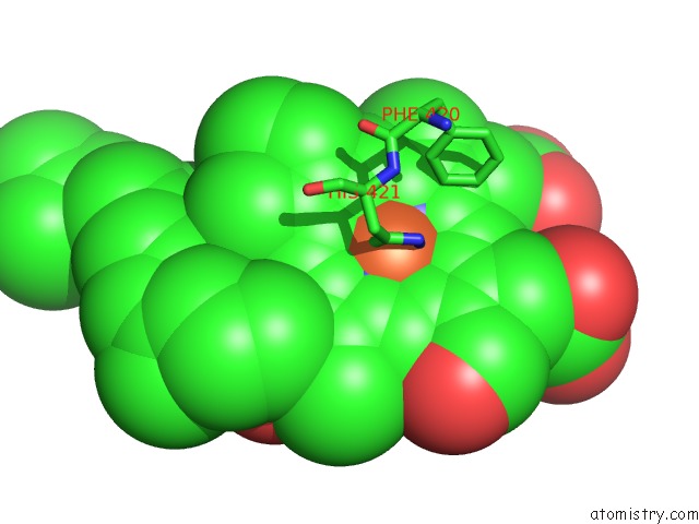
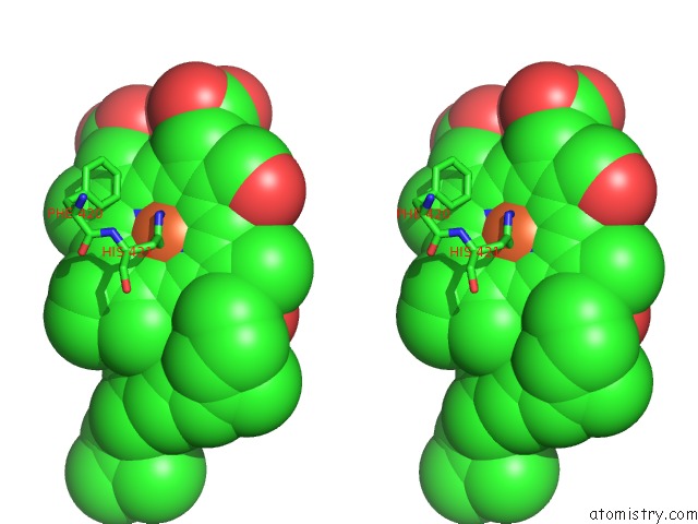
Iron binding site 1 out of 4 in 3omi
Go back to
Iron binding site 1 out
of 4 in the Catalytic Core Subunits (I and II) of Cytochrome C Oxidase From Rhodobacter Sphaeroides with D132A Mutation
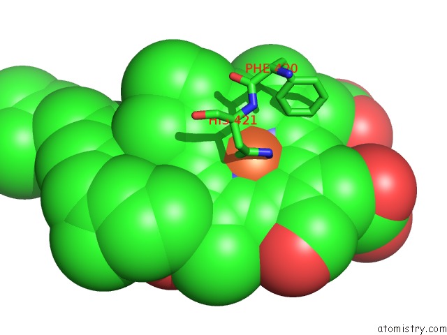
Mono view
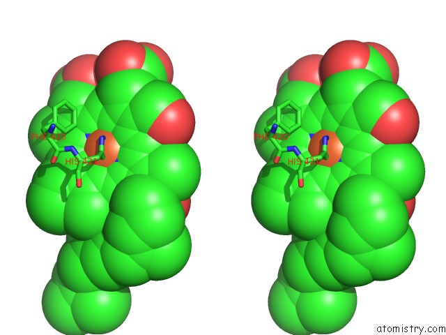
Stereo pair view
Mono view
Stereo pair view
A full contact list of Iron with other atoms in the Fe binding
site number 1 of Catalytic Core Subunits (I and II) of Cytochrome C Oxidase From Rhodobacter Sphaeroides with D132A Mutation within 5.0Å range:
|
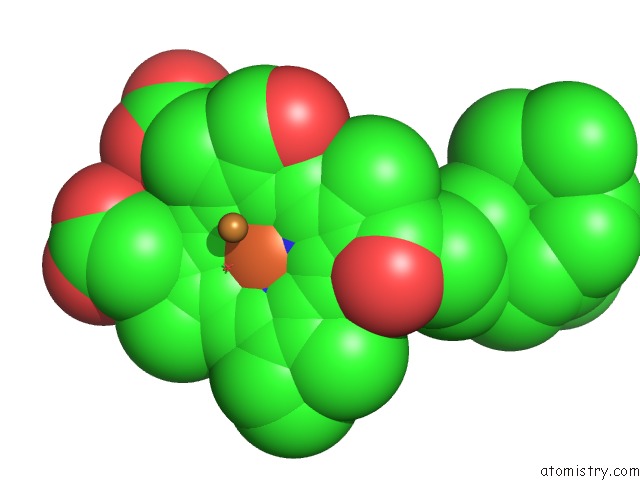
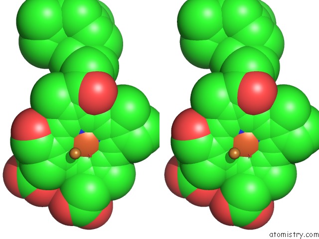
Iron binding site 2 out of 4 in 3omi
Go back to
Iron binding site 2 out
of 4 in the Catalytic Core Subunits (I and II) of Cytochrome C Oxidase From Rhodobacter Sphaeroides with D132A Mutation
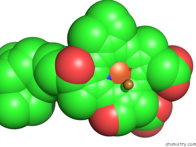
Mono view
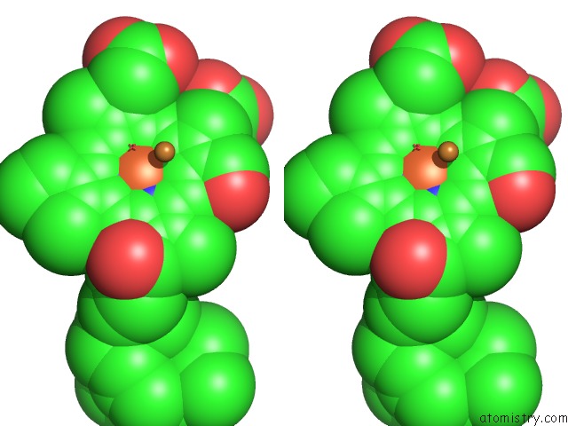
Stereo pair view
Mono view
Stereo pair view
A full contact list of Iron with other atoms in the Fe binding
site number 2 of Catalytic Core Subunits (I and II) of Cytochrome C Oxidase From Rhodobacter Sphaeroides with D132A Mutation within 5.0Å range:
|
Iron binding site 3 out of 4 in 3omi
Go back to
Iron binding site 3 out
of 4 in the Catalytic Core Subunits (I and II) of Cytochrome C Oxidase From Rhodobacter Sphaeroides with D132A Mutation
Mono view
Stereo pair view
Mono view
Stereo pair view
A full contact list of Iron with other atoms in the Fe binding
site number 3 of Catalytic Core Subunits (I and II) of Cytochrome C Oxidase From Rhodobacter Sphaeroides with D132A Mutation within 5.0Å range:
|
Iron binding site 4 out of 4 in 3omi
Go back to
Iron binding site 4 out
of 4 in the Catalytic Core Subunits (I and II) of Cytochrome C Oxidase From Rhodobacter Sphaeroides with D132A Mutation
Mono view
Stereo pair view
Mono view
Stereo pair view
A full contact list of Iron with other atoms in the Fe binding
site number 4 of Catalytic Core Subunits (I and II) of Cytochrome C Oxidase From Rhodobacter Sphaeroides with D132A Mutation within 5.0Å range:
|
Reference:
J.Liu,
L.Qin,
S.Ferguson-Miller.
Crystallographic and Online Spectral Evidence For Role of Conformational Change and Conserved Water in Cytochrome Oxidase Proton Pump. Proc.Natl.Acad.Sci.Usa V. 108 1284 2011.
ISSN: ISSN 0027-8424
PubMed: 21205904
DOI: 10.1073/PNAS.1012846108
Page generated: Sun Aug 4 17:11:58 2024
ISSN: ISSN 0027-8424
PubMed: 21205904
DOI: 10.1073/PNAS.1012846108
Last articles
Fe in 2YXOFe in 2YRS
Fe in 2YXC
Fe in 2YNM
Fe in 2YVJ
Fe in 2YP1
Fe in 2YU2
Fe in 2YU1
Fe in 2YQB
Fe in 2YOO