Iron »
PDB 3om3-3p4q »
3oo3 »
Iron in PDB 3oo3: Crystal Structure of the ORF6* (CYP165D3) Monooxygenase Involved in Teicoplanin Biosynthesis
Protein crystallography data
The structure of Crystal Structure of the ORF6* (CYP165D3) Monooxygenase Involved in Teicoplanin Biosynthesis, PDB code: 3oo3
was solved by
Z.Li,
S.K.Nair,
with X-Ray Crystallography technique. A brief refinement statistics is given in the table below:
| Resolution Low / High (Å) | 25.00 / 2.20 |
| Space group | P 31 |
| Cell size a, b, c (Å), α, β, γ (°) | 74.266, 74.266, 75.467, 90.00, 90.00, 120.00 |
| R / Rfree (%) | 20.1 / 25.8 |
Iron Binding Sites:
The binding sites of Iron atom in the Crystal Structure of the ORF6* (CYP165D3) Monooxygenase Involved in Teicoplanin Biosynthesis
(pdb code 3oo3). This binding sites where shown within
5.0 Angstroms radius around Iron atom.
In total only one binding site of Iron was determined in the Crystal Structure of the ORF6* (CYP165D3) Monooxygenase Involved in Teicoplanin Biosynthesis, PDB code: 3oo3:
In total only one binding site of Iron was determined in the Crystal Structure of the ORF6* (CYP165D3) Monooxygenase Involved in Teicoplanin Biosynthesis, PDB code: 3oo3:
Iron binding site 1 out of 1 in 3oo3
Go back to
Iron binding site 1 out
of 1 in the Crystal Structure of the ORF6* (CYP165D3) Monooxygenase Involved in Teicoplanin Biosynthesis
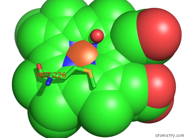
Mono view
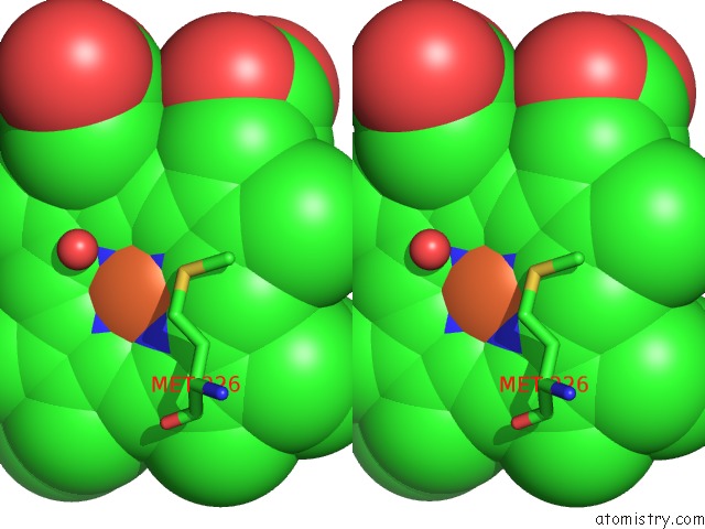
Stereo pair view
Mono view
Stereo pair view
A full contact list of Iron with other atoms in the Fe binding
site number 1 of Crystal Structure of the ORF6* (CYP165D3) Monooxygenase Involved in Teicoplanin Biosynthesis within 5.0Å range:
|
Reference:
Z.Li,
S.G.Rupasinghe,
M.A.Schuler,
S.K.Nair.
Crystal Structure of A Phenol-Coupling P450 Monooxygenase Involved in Teicoplanin Biosynthesis. Proteins V. 79 1728 2011.
ISSN: ISSN 0887-3585
PubMed: 21445994
DOI: 10.1002/PROT.22996
Page generated: Sun Aug 4 17:11:57 2024
ISSN: ISSN 0887-3585
PubMed: 21445994
DOI: 10.1002/PROT.22996
Last articles
Zn in 9J0NZn in 9J0O
Zn in 9J0P
Zn in 9FJX
Zn in 9EKB
Zn in 9C0F
Zn in 9CAH
Zn in 9CH0
Zn in 9CH3
Zn in 9CH1