Iron »
PDB 3om3-3p4q »
3oo4 »
Iron in PDB 3oo4: R-State Human Hemoglobin: Nitriheme Modified at Alpha
Protein crystallography data
The structure of R-State Human Hemoglobin: Nitriheme Modified at Alpha, PDB code: 3oo4
was solved by
J.Yi,
L.M.Thormas,
G.B.Richter-Addo,
with X-Ray Crystallography technique. A brief refinement statistics is given in the table below:
| Resolution Low / High (Å) | 27.20 / 1.90 |
| Space group | P 41 21 2 |
| Cell size a, b, c (Å), α, β, γ (°) | 53.487, 53.487, 190.001, 90.00, 90.00, 90.00 |
| R / Rfree (%) | 20.7 / 26.3 |
Iron Binding Sites:
The binding sites of Iron atom in the R-State Human Hemoglobin: Nitriheme Modified at Alpha
(pdb code 3oo4). This binding sites where shown within
5.0 Angstroms radius around Iron atom.
In total 2 binding sites of Iron where determined in the R-State Human Hemoglobin: Nitriheme Modified at Alpha, PDB code: 3oo4:
Jump to Iron binding site number: 1; 2;
In total 2 binding sites of Iron where determined in the R-State Human Hemoglobin: Nitriheme Modified at Alpha, PDB code: 3oo4:
Jump to Iron binding site number: 1; 2;
Iron binding site 1 out of 2 in 3oo4
Go back to
Iron binding site 1 out
of 2 in the R-State Human Hemoglobin: Nitriheme Modified at Alpha

Mono view
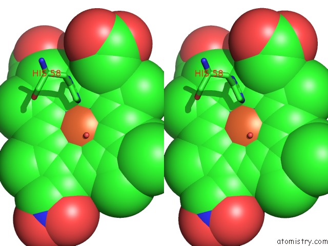
Stereo pair view
Mono view
Stereo pair view
A full contact list of Iron with other atoms in the Fe binding
site number 1 of R-State Human Hemoglobin: Nitriheme Modified at Alpha within 5.0Å range:
|
Iron binding site 2 out of 2 in 3oo4
Go back to
Iron binding site 2 out
of 2 in the R-State Human Hemoglobin: Nitriheme Modified at Alpha
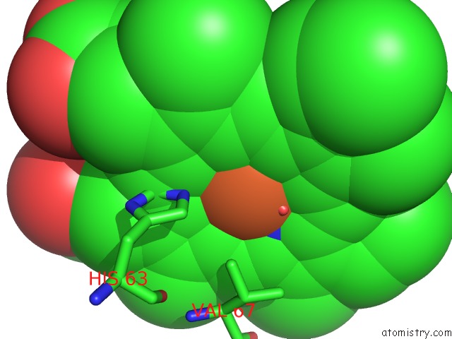
Mono view

Stereo pair view
Mono view
Stereo pair view
A full contact list of Iron with other atoms in the Fe binding
site number 2 of R-State Human Hemoglobin: Nitriheme Modified at Alpha within 5.0Å range:
|
Reference:
J.Yi,
L.M.Thomas,
F.N.Musayev,
M.K.Safo,
G.B.Richter-Addo.
Crystallographic Trapping of Heme Loss Intermediates During the Nitrite-Induced Degradation of Human Hemoglobin. Biochemistry V. 50 8323 2011.
ISSN: ISSN 0006-2960
PubMed: 21863786
DOI: 10.1021/BI2009322
Page generated: Sun Aug 4 17:11:57 2024
ISSN: ISSN 0006-2960
PubMed: 21863786
DOI: 10.1021/BI2009322
Last articles
Cl in 5V8SCl in 5V8P
Cl in 5V9J
Cl in 5V86
Cl in 5V7G
Cl in 5V8G
Cl in 5V71
Cl in 5V6I
Cl in 5V7E
Cl in 5V72