Iron »
PDB 3om3-3p4q »
3p3h »
Iron in PDB 3p3h: Human Carbonic Anhydrase II in Complex with P-(5-Ferrocenyl-1H-1,2,3- Triazol-1-Yl)Benzenesulfonamide
Enzymatic activity of Human Carbonic Anhydrase II in Complex with P-(5-Ferrocenyl-1H-1,2,3- Triazol-1-Yl)Benzenesulfonamide
All present enzymatic activity of Human Carbonic Anhydrase II in Complex with P-(5-Ferrocenyl-1H-1,2,3- Triazol-1-Yl)Benzenesulfonamide:
4.2.1.1;
4.2.1.1;
Protein crystallography data
The structure of Human Carbonic Anhydrase II in Complex with P-(5-Ferrocenyl-1H-1,2,3- Triazol-1-Yl)Benzenesulfonamide, PDB code: 3p3h
was solved by
A.J.Salmon,
with X-Ray Crystallography technique. A brief refinement statistics is given in the table below:
| Resolution Low / High (Å) | 22.92 / 1.50 |
| Space group | P 1 21 1 |
| Cell size a, b, c (Å), α, β, γ (°) | 42.456, 41.574, 72.226, 90.00, 104.81, 90.00 |
| R / Rfree (%) | 17.6 / 20.1 |
Other elements in 3p3h:
The structure of Human Carbonic Anhydrase II in Complex with P-(5-Ferrocenyl-1H-1,2,3- Triazol-1-Yl)Benzenesulfonamide also contains other interesting chemical elements:
| Zinc | (Zn) | 1 atom |
Iron Binding Sites:
The binding sites of Iron atom in the Human Carbonic Anhydrase II in Complex with P-(5-Ferrocenyl-1H-1,2,3- Triazol-1-Yl)Benzenesulfonamide
(pdb code 3p3h). This binding sites where shown within
5.0 Angstroms radius around Iron atom.
In total only one binding site of Iron was determined in the Human Carbonic Anhydrase II in Complex with P-(5-Ferrocenyl-1H-1,2,3- Triazol-1-Yl)Benzenesulfonamide, PDB code: 3p3h:
In total only one binding site of Iron was determined in the Human Carbonic Anhydrase II in Complex with P-(5-Ferrocenyl-1H-1,2,3- Triazol-1-Yl)Benzenesulfonamide, PDB code: 3p3h:
Iron binding site 1 out of 1 in 3p3h
Go back to
Iron binding site 1 out
of 1 in the Human Carbonic Anhydrase II in Complex with P-(5-Ferrocenyl-1H-1,2,3- Triazol-1-Yl)Benzenesulfonamide
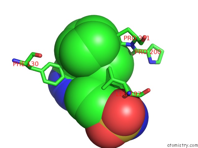
Mono view
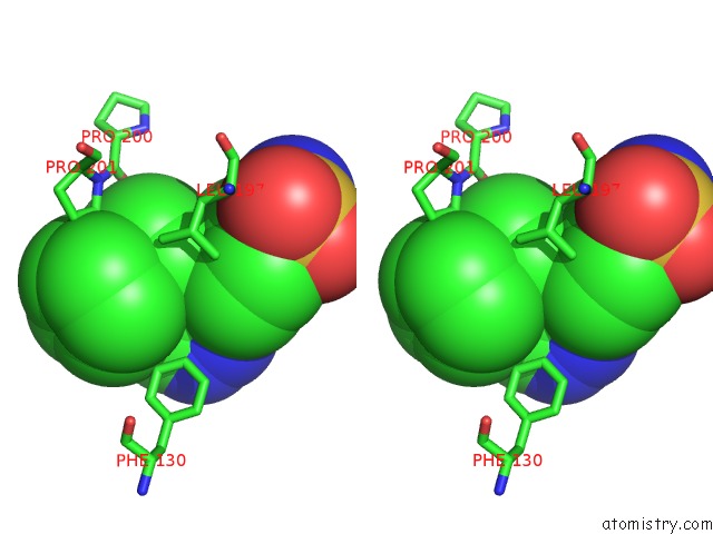
Stereo pair view
Mono view
Stereo pair view
A full contact list of Iron with other atoms in the Fe binding
site number 1 of Human Carbonic Anhydrase II in Complex with P-(5-Ferrocenyl-1H-1,2,3- Triazol-1-Yl)Benzenesulfonamide within 5.0Å range:
|
Reference:
A.J.Salmon,
M.L.Williams,
A.Hofmann,
S.A.Poulsen.
Protein Crystal Structures with Ferrocene and Ruthenocene-Based Enzyme Inhibitors. Chem.Commun.(Camb.) V. 48 2328 2012.
ISSN: ISSN 1359-7345
PubMed: 22258283
DOI: 10.1039/C2CC15625C
Page generated: Sun Aug 4 17:22:02 2024
ISSN: ISSN 1359-7345
PubMed: 22258283
DOI: 10.1039/C2CC15625C
Last articles
Zn in 9MJ5Zn in 9HNW
Zn in 9G0L
Zn in 9FNE
Zn in 9DZN
Zn in 9E0I
Zn in 9D32
Zn in 9DAK
Zn in 8ZXC
Zn in 8ZUF