Iron »
PDB 3p4r-3pcn »
3pcf »
Iron in PDB 3pcf: Structure of Protocatechuate 3,4-Dioxygenase Complexed with 3-Fluro-4- Hydroxybenzoate
Enzymatic activity of Structure of Protocatechuate 3,4-Dioxygenase Complexed with 3-Fluro-4- Hydroxybenzoate
All present enzymatic activity of Structure of Protocatechuate 3,4-Dioxygenase Complexed with 3-Fluro-4- Hydroxybenzoate:
1.13.11.3;
1.13.11.3;
Protein crystallography data
The structure of Structure of Protocatechuate 3,4-Dioxygenase Complexed with 3-Fluro-4- Hydroxybenzoate, PDB code: 3pcf
was solved by
A.M.Orville,
N.Elango,
J.D.Lipscomb,
D.H.Ohlendorf,
with X-Ray Crystallography technique. A brief refinement statistics is given in the table below:
| Resolution Low / High (Å) | 6.00 / 2.15 |
| Space group | I 1 2 1 |
| Cell size a, b, c (Å), α, β, γ (°) | 196.030, 127.220, 133.700, 90.00, 97.70, 90.00 |
| R / Rfree (%) | n/a / n/a |
Other elements in 3pcf:
The structure of Structure of Protocatechuate 3,4-Dioxygenase Complexed with 3-Fluro-4- Hydroxybenzoate also contains other interesting chemical elements:
| Fluorine | (F) | 12 atoms |
Iron Binding Sites:
The binding sites of Iron atom in the Structure of Protocatechuate 3,4-Dioxygenase Complexed with 3-Fluro-4- Hydroxybenzoate
(pdb code 3pcf). This binding sites where shown within
5.0 Angstroms radius around Iron atom.
In total 6 binding sites of Iron where determined in the Structure of Protocatechuate 3,4-Dioxygenase Complexed with 3-Fluro-4- Hydroxybenzoate, PDB code: 3pcf:
Jump to Iron binding site number: 1; 2; 3; 4; 5; 6;
In total 6 binding sites of Iron where determined in the Structure of Protocatechuate 3,4-Dioxygenase Complexed with 3-Fluro-4- Hydroxybenzoate, PDB code: 3pcf:
Jump to Iron binding site number: 1; 2; 3; 4; 5; 6;
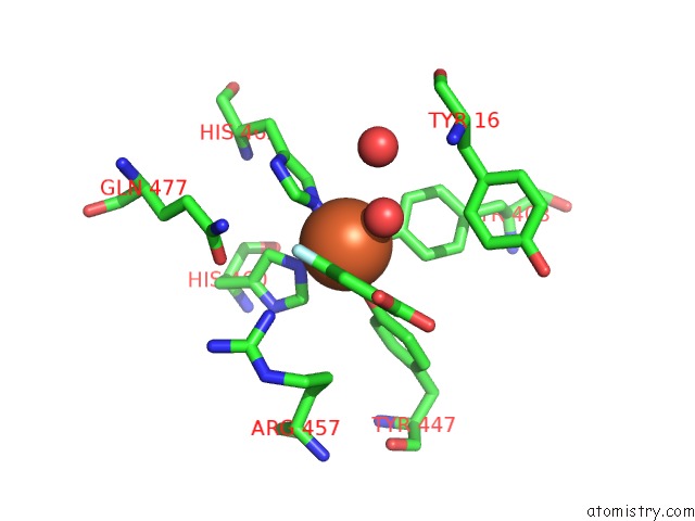
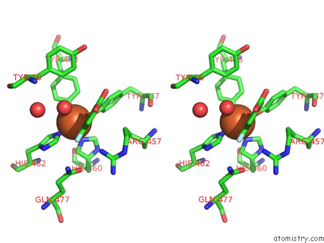
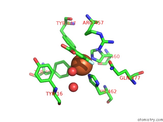
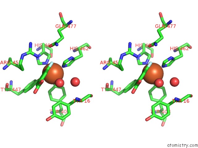
Iron binding site 1 out of 6 in 3pcf
Go back to
Iron binding site 1 out
of 6 in the Structure of Protocatechuate 3,4-Dioxygenase Complexed with 3-Fluro-4- Hydroxybenzoate
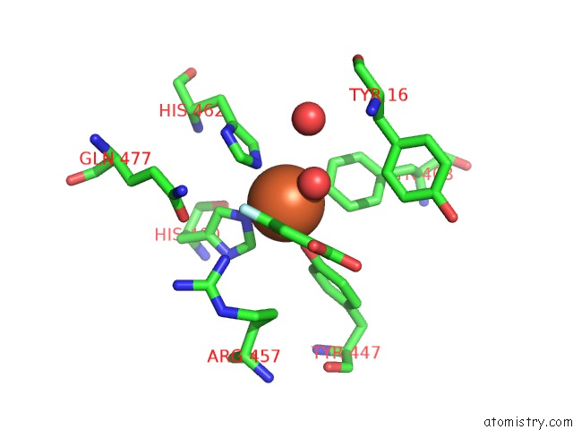
Mono view
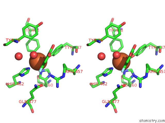
Stereo pair view
Mono view
Stereo pair view
A full contact list of Iron with other atoms in the Fe binding
site number 1 of Structure of Protocatechuate 3,4-Dioxygenase Complexed with 3-Fluro-4- Hydroxybenzoate within 5.0Å range:
|
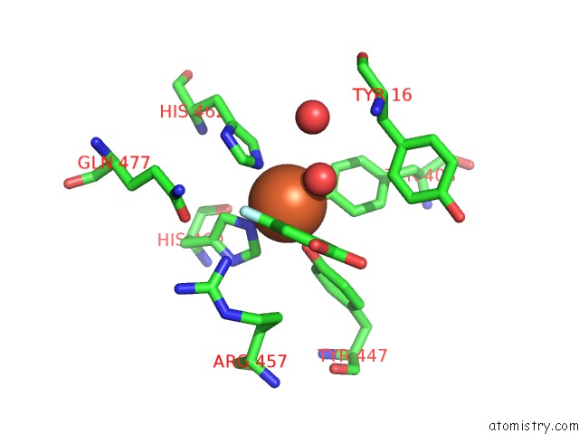
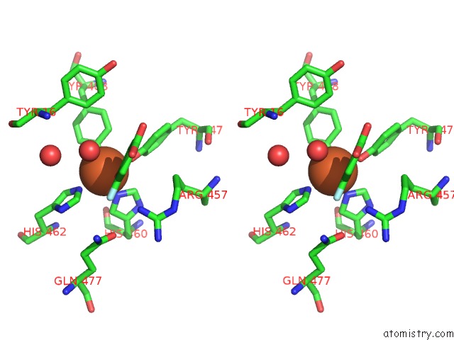
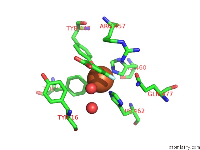
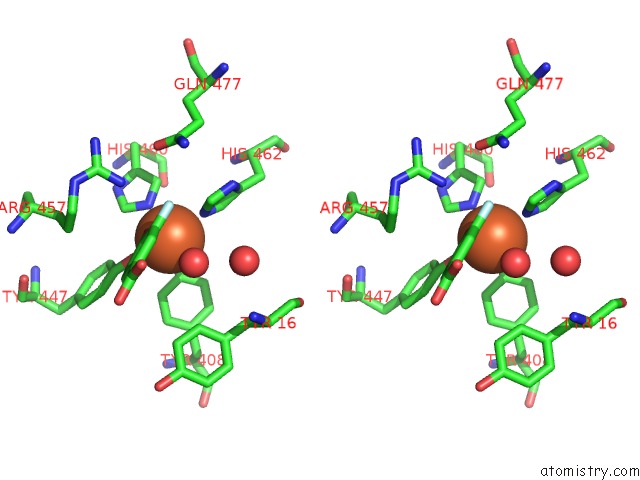
Iron binding site 2 out of 6 in 3pcf
Go back to
Iron binding site 2 out
of 6 in the Structure of Protocatechuate 3,4-Dioxygenase Complexed with 3-Fluro-4- Hydroxybenzoate
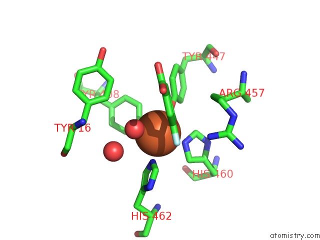
Mono view
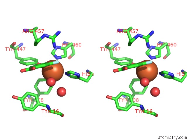
Stereo pair view
Mono view
Stereo pair view
A full contact list of Iron with other atoms in the Fe binding
site number 2 of Structure of Protocatechuate 3,4-Dioxygenase Complexed with 3-Fluro-4- Hydroxybenzoate within 5.0Å range:
|
Iron binding site 3 out of 6 in 3pcf
Go back to
Iron binding site 3 out
of 6 in the Structure of Protocatechuate 3,4-Dioxygenase Complexed with 3-Fluro-4- Hydroxybenzoate
Mono view
Stereo pair view
Mono view
Stereo pair view
A full contact list of Iron with other atoms in the Fe binding
site number 3 of Structure of Protocatechuate 3,4-Dioxygenase Complexed with 3-Fluro-4- Hydroxybenzoate within 5.0Å range:
|
Iron binding site 4 out of 6 in 3pcf
Go back to
Iron binding site 4 out
of 6 in the Structure of Protocatechuate 3,4-Dioxygenase Complexed with 3-Fluro-4- Hydroxybenzoate
Mono view
Stereo pair view
Mono view
Stereo pair view
A full contact list of Iron with other atoms in the Fe binding
site number 4 of Structure of Protocatechuate 3,4-Dioxygenase Complexed with 3-Fluro-4- Hydroxybenzoate within 5.0Å range:
|
Iron binding site 5 out of 6 in 3pcf
Go back to
Iron binding site 5 out
of 6 in the Structure of Protocatechuate 3,4-Dioxygenase Complexed with 3-Fluro-4- Hydroxybenzoate
Mono view
Stereo pair view
Mono view
Stereo pair view
A full contact list of Iron with other atoms in the Fe binding
site number 5 of Structure of Protocatechuate 3,4-Dioxygenase Complexed with 3-Fluro-4- Hydroxybenzoate within 5.0Å range:
|
Iron binding site 6 out of 6 in 3pcf
Go back to
Iron binding site 6 out
of 6 in the Structure of Protocatechuate 3,4-Dioxygenase Complexed with 3-Fluro-4- Hydroxybenzoate
Mono view
Stereo pair view
Mono view
Stereo pair view
A full contact list of Iron with other atoms in the Fe binding
site number 6 of Structure of Protocatechuate 3,4-Dioxygenase Complexed with 3-Fluro-4- Hydroxybenzoate within 5.0Å range:
|
Reference:
A.M.Orville,
N.Elango,
J.D.Lipscomb,
D.H.Ohlendorf.
Structures of Competitive Inhibitor Complexes of Protocatechuate 3,4-Dioxygenase: Multiple Exogenous Ligand Binding Orientations Within the Active Site. Biochemistry V. 36 10039 1997.
ISSN: ISSN 0006-2960
PubMed: 9254599
DOI: 10.1021/BI970468N
Page generated: Sun Aug 4 17:49:34 2024
ISSN: ISSN 0006-2960
PubMed: 9254599
DOI: 10.1021/BI970468N
Last articles
Zn in 9JYWZn in 9IR4
Zn in 9IR3
Zn in 9GMX
Zn in 9GMW
Zn in 9JEJ
Zn in 9ERF
Zn in 9ERE
Zn in 9EGV
Zn in 9EGW