Iron »
PDB 3p4r-3pcn »
3pcj »
Iron in PDB 3pcj: Structure of Protocatechuate 3,4-Dioxygenase Complexed with 2- Hydroxyisonicotinic Acid N-Oxide
Enzymatic activity of Structure of Protocatechuate 3,4-Dioxygenase Complexed with 2- Hydroxyisonicotinic Acid N-Oxide
All present enzymatic activity of Structure of Protocatechuate 3,4-Dioxygenase Complexed with 2- Hydroxyisonicotinic Acid N-Oxide:
1.13.11.3;
1.13.11.3;
Protein crystallography data
The structure of Structure of Protocatechuate 3,4-Dioxygenase Complexed with 2- Hydroxyisonicotinic Acid N-Oxide, PDB code: 3pcj
was solved by
A.M.Orville,
J.D.Lipscomb,
D.H.Ohlendorf,
with X-Ray Crystallography technique. A brief refinement statistics is given in the table below:
| Resolution Low / High (Å) | 6.00 / 2.13 |
| Space group | I 1 2 1 |
| Cell size a, b, c (Å), α, β, γ (°) | 196.200, 127.100, 133.700, 90.00, 97.70, 90.00 |
| R / Rfree (%) | n/a / n/a |
Iron Binding Sites:
The binding sites of Iron atom in the Structure of Protocatechuate 3,4-Dioxygenase Complexed with 2- Hydroxyisonicotinic Acid N-Oxide
(pdb code 3pcj). This binding sites where shown within
5.0 Angstroms radius around Iron atom.
In total 6 binding sites of Iron where determined in the Structure of Protocatechuate 3,4-Dioxygenase Complexed with 2- Hydroxyisonicotinic Acid N-Oxide, PDB code: 3pcj:
Jump to Iron binding site number: 1; 2; 3; 4; 5; 6;
In total 6 binding sites of Iron where determined in the Structure of Protocatechuate 3,4-Dioxygenase Complexed with 2- Hydroxyisonicotinic Acid N-Oxide, PDB code: 3pcj:
Jump to Iron binding site number: 1; 2; 3; 4; 5; 6;
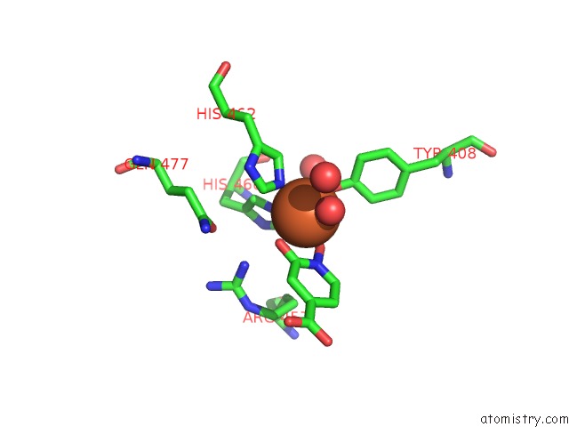
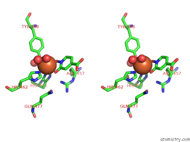
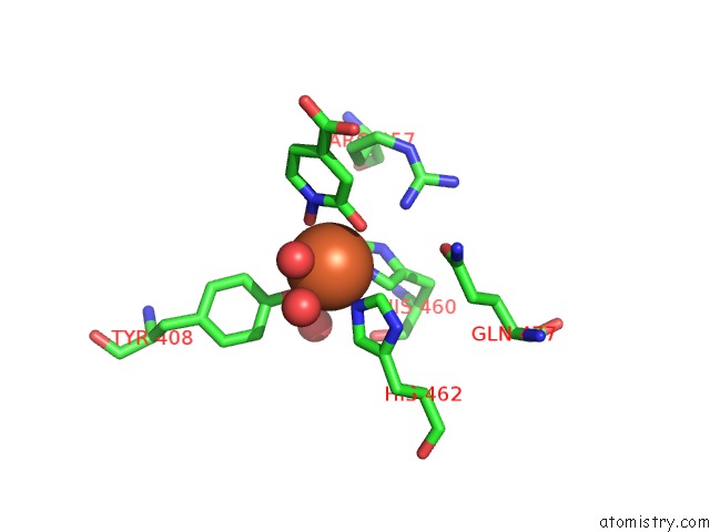
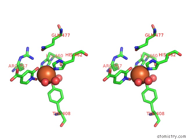
Iron binding site 1 out of 6 in 3pcj
Go back to
Iron binding site 1 out
of 6 in the Structure of Protocatechuate 3,4-Dioxygenase Complexed with 2- Hydroxyisonicotinic Acid N-Oxide
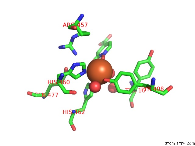
Mono view
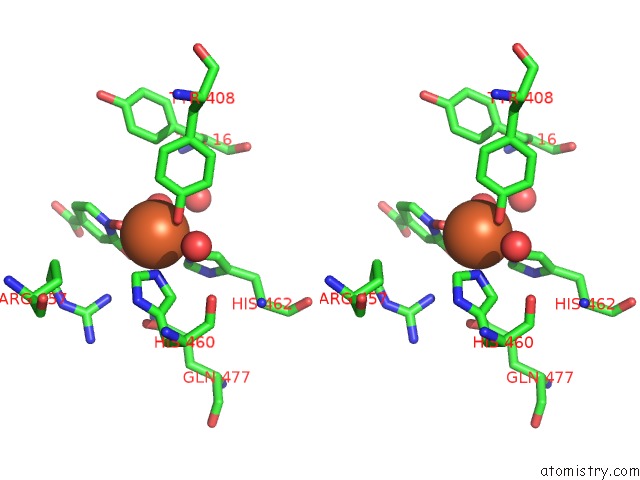
Stereo pair view
Mono view
Stereo pair view
A full contact list of Iron with other atoms in the Fe binding
site number 1 of Structure of Protocatechuate 3,4-Dioxygenase Complexed with 2- Hydroxyisonicotinic Acid N-Oxide within 5.0Å range:
|
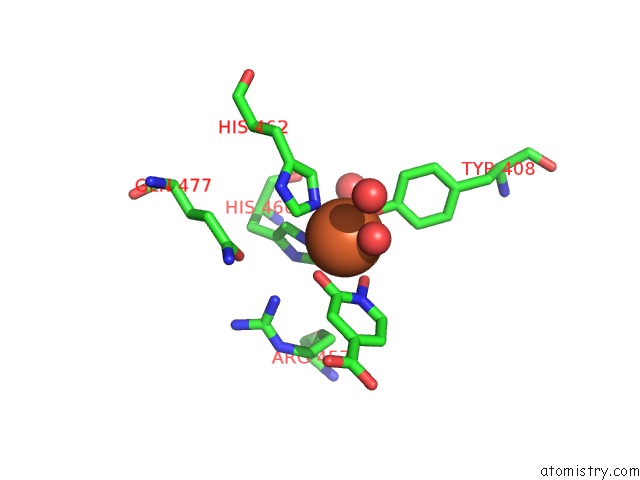
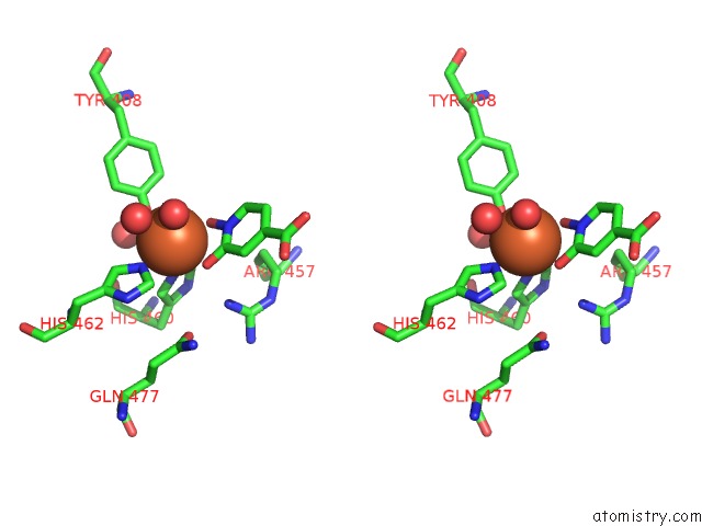
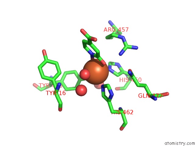
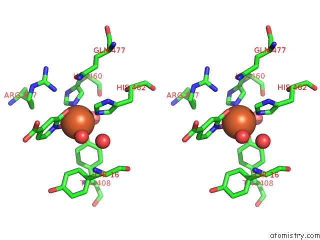
Iron binding site 2 out of 6 in 3pcj
Go back to
Iron binding site 2 out
of 6 in the Structure of Protocatechuate 3,4-Dioxygenase Complexed with 2- Hydroxyisonicotinic Acid N-Oxide
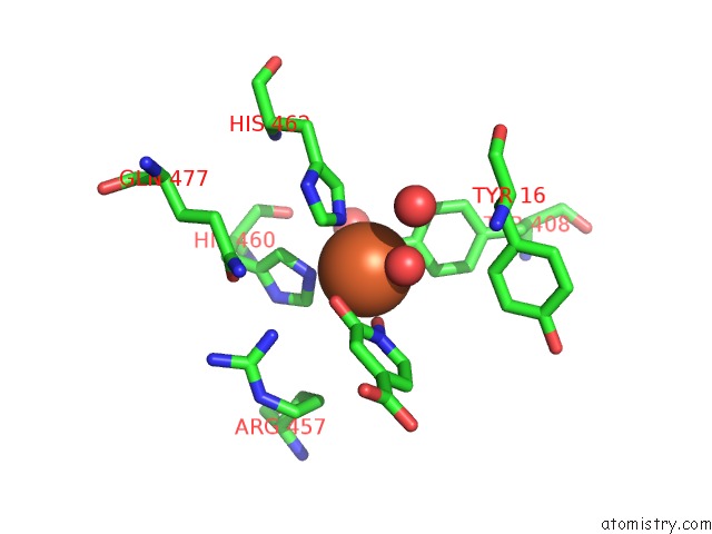
Mono view
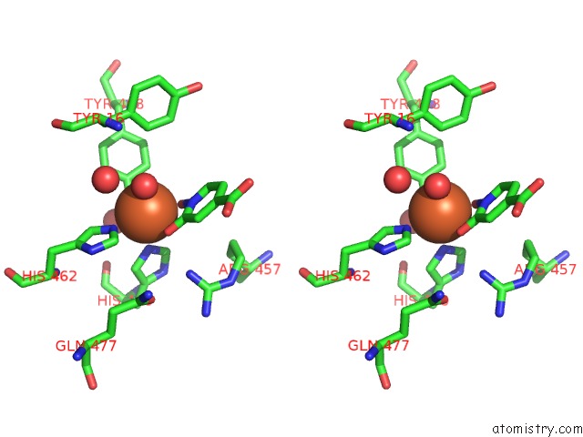
Stereo pair view
Mono view
Stereo pair view
A full contact list of Iron with other atoms in the Fe binding
site number 2 of Structure of Protocatechuate 3,4-Dioxygenase Complexed with 2- Hydroxyisonicotinic Acid N-Oxide within 5.0Å range:
|
Iron binding site 3 out of 6 in 3pcj
Go back to
Iron binding site 3 out
of 6 in the Structure of Protocatechuate 3,4-Dioxygenase Complexed with 2- Hydroxyisonicotinic Acid N-Oxide
Mono view
Stereo pair view
Mono view
Stereo pair view
A full contact list of Iron with other atoms in the Fe binding
site number 3 of Structure of Protocatechuate 3,4-Dioxygenase Complexed with 2- Hydroxyisonicotinic Acid N-Oxide within 5.0Å range:
|
Iron binding site 4 out of 6 in 3pcj
Go back to
Iron binding site 4 out
of 6 in the Structure of Protocatechuate 3,4-Dioxygenase Complexed with 2- Hydroxyisonicotinic Acid N-Oxide
Mono view
Stereo pair view
Mono view
Stereo pair view
A full contact list of Iron with other atoms in the Fe binding
site number 4 of Structure of Protocatechuate 3,4-Dioxygenase Complexed with 2- Hydroxyisonicotinic Acid N-Oxide within 5.0Å range:
|
Iron binding site 5 out of 6 in 3pcj
Go back to
Iron binding site 5 out
of 6 in the Structure of Protocatechuate 3,4-Dioxygenase Complexed with 2- Hydroxyisonicotinic Acid N-Oxide
Mono view
Stereo pair view
Mono view
Stereo pair view
A full contact list of Iron with other atoms in the Fe binding
site number 5 of Structure of Protocatechuate 3,4-Dioxygenase Complexed with 2- Hydroxyisonicotinic Acid N-Oxide within 5.0Å range:
|
Iron binding site 6 out of 6 in 3pcj
Go back to
Iron binding site 6 out
of 6 in the Structure of Protocatechuate 3,4-Dioxygenase Complexed with 2- Hydroxyisonicotinic Acid N-Oxide
Mono view
Stereo pair view
Mono view
Stereo pair view
A full contact list of Iron with other atoms in the Fe binding
site number 6 of Structure of Protocatechuate 3,4-Dioxygenase Complexed with 2- Hydroxyisonicotinic Acid N-Oxide within 5.0Å range:
|
Reference:
A.M.Orville,
J.D.Lipscomb,
D.H.Ohlendorf.
Crystal Structures of Substrate and Substrate Analog Complexes of Protocatechuate 3,4-Dioxygenase: Endogenous FE3+ Ligand Displacement in Response to Substrate Binding. Biochemistry V. 36 10052 1997.
ISSN: ISSN 0006-2960
PubMed: 9254600
DOI: 10.1021/BI970469F
Page generated: Sun Aug 4 17:52:18 2024
ISSN: ISSN 0006-2960
PubMed: 9254600
DOI: 10.1021/BI970469F
Last articles
Zn in 9J0NZn in 9J0O
Zn in 9J0P
Zn in 9FJX
Zn in 9EKB
Zn in 9C0F
Zn in 9CAH
Zn in 9CH0
Zn in 9CH3
Zn in 9CH1