Iron »
PDB 3pur-3qjs »
3pxw »
Iron in PDB 3pxw: Crystal Structure of Ferrous No Adduct of Maug in Complex with Pre- Methylamine Dehydrogenase
Enzymatic activity of Crystal Structure of Ferrous No Adduct of Maug in Complex with Pre- Methylamine Dehydrogenase
All present enzymatic activity of Crystal Structure of Ferrous No Adduct of Maug in Complex with Pre- Methylamine Dehydrogenase:
1.4.99.3;
1.4.99.3;
Protein crystallography data
The structure of Crystal Structure of Ferrous No Adduct of Maug in Complex with Pre- Methylamine Dehydrogenase, PDB code: 3pxw
was solved by
E.T.Yukl,
B.R.Goblirsch,
C.M.Wilmot,
with X-Ray Crystallography technique. A brief refinement statistics is given in the table below:
| Resolution Low / High (Å) | 44.49 / 2.11 |
| Space group | P 1 |
| Cell size a, b, c (Å), α, β, γ (°) | 55.530, 83.520, 107.780, 109.94, 91.54, 105.78 |
| R / Rfree (%) | 18.9 / 24.2 |
Other elements in 3pxw:
The structure of Crystal Structure of Ferrous No Adduct of Maug in Complex with Pre- Methylamine Dehydrogenase also contains other interesting chemical elements:
| Calcium | (Ca) | 2 atoms |
| Sodium | (Na) | 2 atoms |
Iron Binding Sites:
The binding sites of Iron atom in the Crystal Structure of Ferrous No Adduct of Maug in Complex with Pre- Methylamine Dehydrogenase
(pdb code 3pxw). This binding sites where shown within
5.0 Angstroms radius around Iron atom.
In total 4 binding sites of Iron where determined in the Crystal Structure of Ferrous No Adduct of Maug in Complex with Pre- Methylamine Dehydrogenase, PDB code: 3pxw:
Jump to Iron binding site number: 1; 2; 3; 4;
In total 4 binding sites of Iron where determined in the Crystal Structure of Ferrous No Adduct of Maug in Complex with Pre- Methylamine Dehydrogenase, PDB code: 3pxw:
Jump to Iron binding site number: 1; 2; 3; 4;
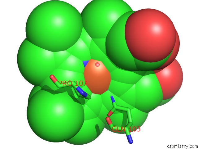
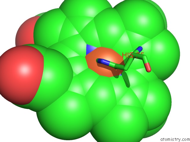
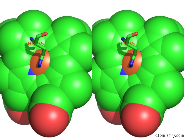
Iron binding site 1 out of 4 in 3pxw
Go back to
Iron binding site 1 out
of 4 in the Crystal Structure of Ferrous No Adduct of Maug in Complex with Pre- Methylamine Dehydrogenase
Mono view
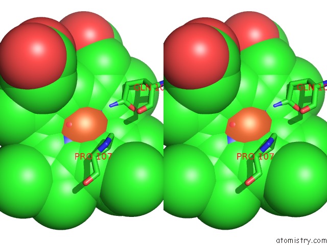
Stereo pair view
Mono view
Stereo pair view
A full contact list of Iron with other atoms in the Fe binding
site number 1 of Crystal Structure of Ferrous No Adduct of Maug in Complex with Pre- Methylamine Dehydrogenase within 5.0Å range:
|
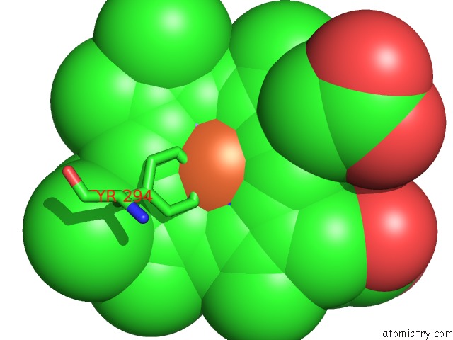
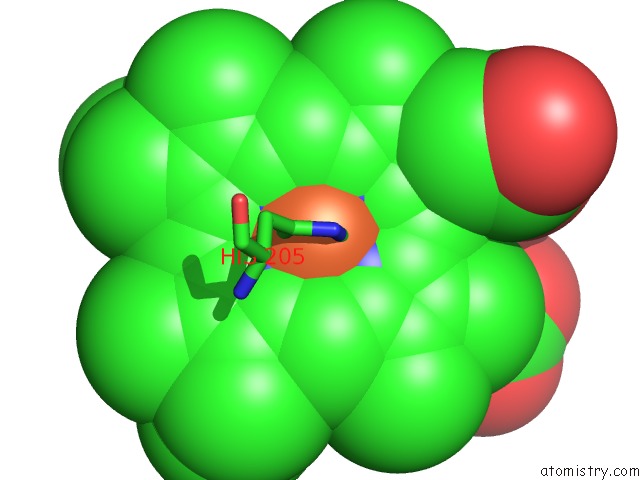
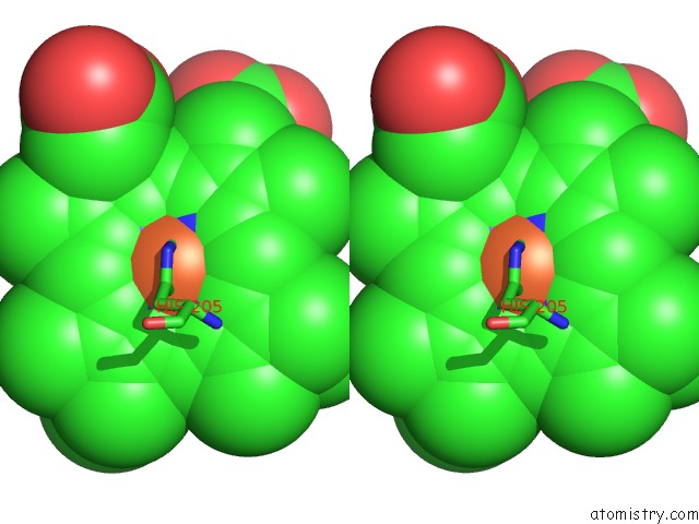
Iron binding site 2 out of 4 in 3pxw
Go back to
Iron binding site 2 out
of 4 in the Crystal Structure of Ferrous No Adduct of Maug in Complex with Pre- Methylamine Dehydrogenase
Mono view
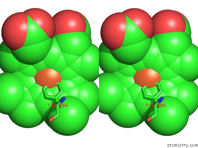
Stereo pair view
Mono view
Stereo pair view
A full contact list of Iron with other atoms in the Fe binding
site number 2 of Crystal Structure of Ferrous No Adduct of Maug in Complex with Pre- Methylamine Dehydrogenase within 5.0Å range:
|
Iron binding site 3 out of 4 in 3pxw
Go back to
Iron binding site 3 out
of 4 in the Crystal Structure of Ferrous No Adduct of Maug in Complex with Pre- Methylamine Dehydrogenase
Mono view
Stereo pair view
Mono view
Stereo pair view
A full contact list of Iron with other atoms in the Fe binding
site number 3 of Crystal Structure of Ferrous No Adduct of Maug in Complex with Pre- Methylamine Dehydrogenase within 5.0Å range:
|
Iron binding site 4 out of 4 in 3pxw
Go back to
Iron binding site 4 out
of 4 in the Crystal Structure of Ferrous No Adduct of Maug in Complex with Pre- Methylamine Dehydrogenase
Mono view
Stereo pair view
Mono view
Stereo pair view
A full contact list of Iron with other atoms in the Fe binding
site number 4 of Crystal Structure of Ferrous No Adduct of Maug in Complex with Pre- Methylamine Dehydrogenase within 5.0Å range:
|
Reference:
E.T.Yukl,
B.R.Goblirsch,
V.L.Davidson,
C.M.Wilmot.
Crystal Structures of Co and No Adducts of Maug in Complex with Pre-Methylamine Dehydrogenase: Implications For the Mechanism of Dioxygen Activation. Biochemistry V. 50 2931 2011.
ISSN: ISSN 0006-2960
PubMed: 21355604
DOI: 10.1021/BI200023N
Page generated: Sun Aug 4 18:29:30 2024
ISSN: ISSN 0006-2960
PubMed: 21355604
DOI: 10.1021/BI200023N
Last articles
Zn in 9J0NZn in 9J0O
Zn in 9J0P
Zn in 9FJX
Zn in 9EKB
Zn in 9C0F
Zn in 9CAH
Zn in 9CH0
Zn in 9CH3
Zn in 9CH1