Iron »
PDB 3qjt-3r0g »
3qu8 »
Iron in PDB 3qu8: Crystal Structure of A Human Cytochrome P450 2B6 (Y226H/K262R) in Complex with the Inhibitor 4-(4-Nitrobenzyl)Pyridine.
Enzymatic activity of Crystal Structure of A Human Cytochrome P450 2B6 (Y226H/K262R) in Complex with the Inhibitor 4-(4-Nitrobenzyl)Pyridine.
All present enzymatic activity of Crystal Structure of A Human Cytochrome P450 2B6 (Y226H/K262R) in Complex with the Inhibitor 4-(4-Nitrobenzyl)Pyridine.:
1.14.14.1;
1.14.14.1;
Protein crystallography data
The structure of Crystal Structure of A Human Cytochrome P450 2B6 (Y226H/K262R) in Complex with the Inhibitor 4-(4-Nitrobenzyl)Pyridine., PDB code: 3qu8
was solved by
M.B.Shah,
J.Pascual,
C.D.Stout,
J.R.Halpert,
with X-Ray Crystallography technique. A brief refinement statistics is given in the table below:
| Resolution Low / High (Å) | 50.51 / 2.80 |
| Space group | P 32 |
| Cell size a, b, c (Å), α, β, γ (°) | 101.880, 101.880, 299.509, 90.00, 90.00, 120.00 |
| R / Rfree (%) | 21.8 / 25.9 |
Iron Binding Sites:
The binding sites of Iron atom in the Crystal Structure of A Human Cytochrome P450 2B6 (Y226H/K262R) in Complex with the Inhibitor 4-(4-Nitrobenzyl)Pyridine.
(pdb code 3qu8). This binding sites where shown within
5.0 Angstroms radius around Iron atom.
In total 6 binding sites of Iron where determined in the Crystal Structure of A Human Cytochrome P450 2B6 (Y226H/K262R) in Complex with the Inhibitor 4-(4-Nitrobenzyl)Pyridine., PDB code: 3qu8:
Jump to Iron binding site number: 1; 2; 3; 4; 5; 6;
In total 6 binding sites of Iron where determined in the Crystal Structure of A Human Cytochrome P450 2B6 (Y226H/K262R) in Complex with the Inhibitor 4-(4-Nitrobenzyl)Pyridine., PDB code: 3qu8:
Jump to Iron binding site number: 1; 2; 3; 4; 5; 6;
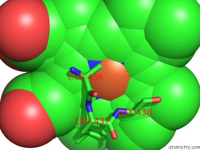
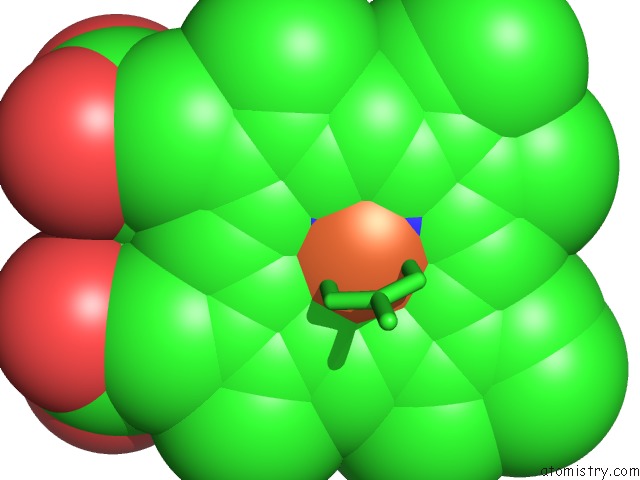
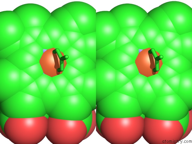
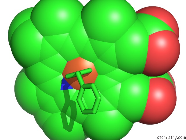
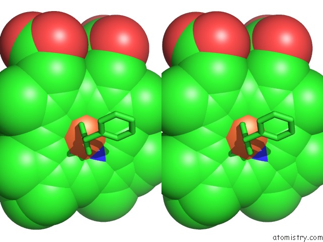
Iron binding site 1 out of 6 in 3qu8
Go back to
Iron binding site 1 out
of 6 in the Crystal Structure of A Human Cytochrome P450 2B6 (Y226H/K262R) in Complex with the Inhibitor 4-(4-Nitrobenzyl)Pyridine.
Mono view
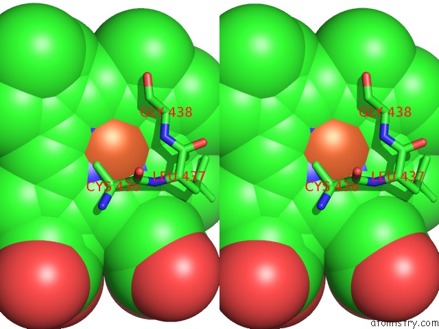
Stereo pair view
Mono view
Stereo pair view
A full contact list of Iron with other atoms in the Fe binding
site number 1 of Crystal Structure of A Human Cytochrome P450 2B6 (Y226H/K262R) in Complex with the Inhibitor 4-(4-Nitrobenzyl)Pyridine. within 5.0Å range:
|
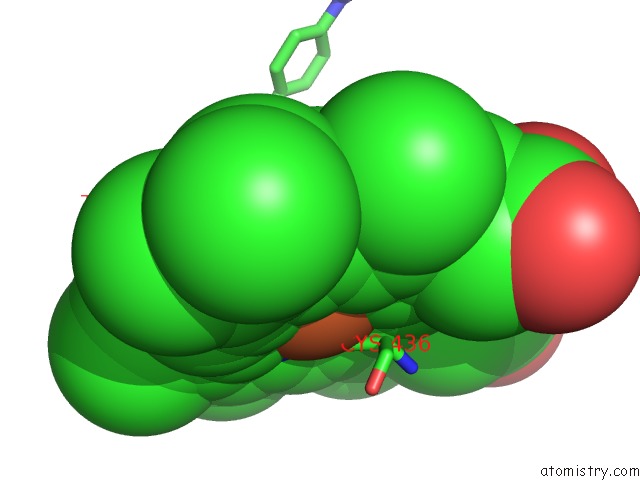
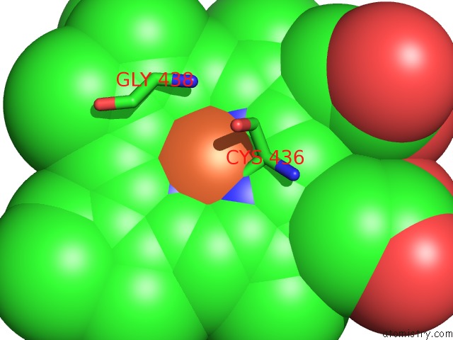
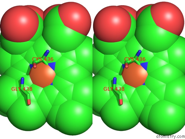
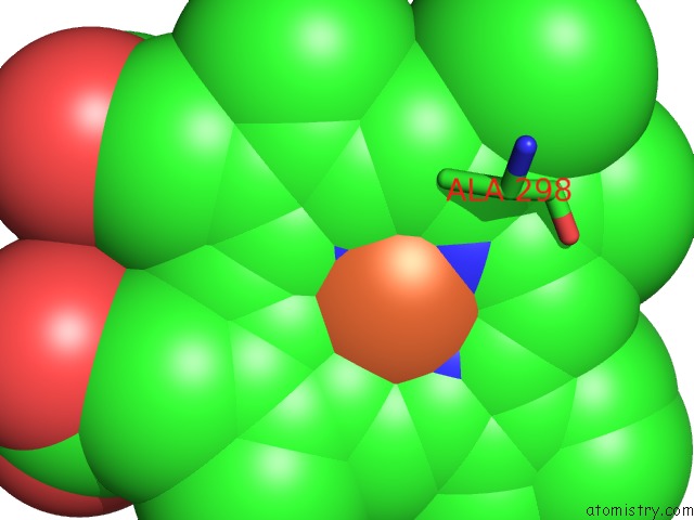
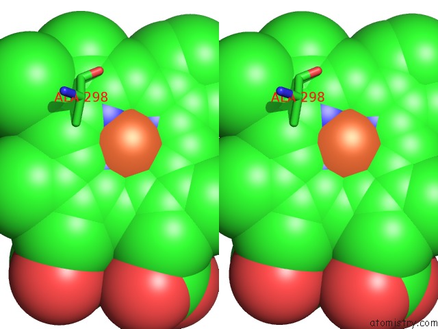
Iron binding site 2 out of 6 in 3qu8
Go back to
Iron binding site 2 out
of 6 in the Crystal Structure of A Human Cytochrome P450 2B6 (Y226H/K262R) in Complex with the Inhibitor 4-(4-Nitrobenzyl)Pyridine.
Mono view
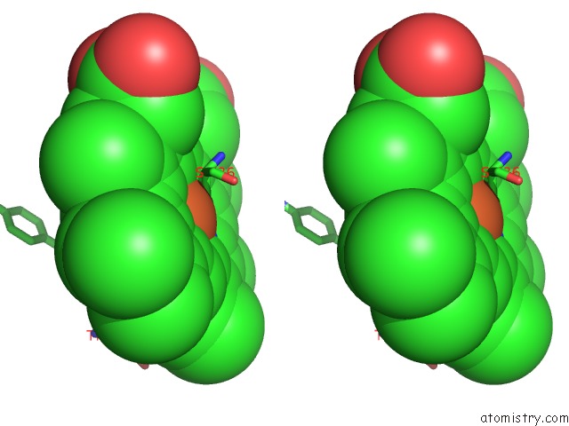
Stereo pair view
Mono view
Stereo pair view
A full contact list of Iron with other atoms in the Fe binding
site number 2 of Crystal Structure of A Human Cytochrome P450 2B6 (Y226H/K262R) in Complex with the Inhibitor 4-(4-Nitrobenzyl)Pyridine. within 5.0Å range:
|
Iron binding site 3 out of 6 in 3qu8
Go back to
Iron binding site 3 out
of 6 in the Crystal Structure of A Human Cytochrome P450 2B6 (Y226H/K262R) in Complex with the Inhibitor 4-(4-Nitrobenzyl)Pyridine.
Mono view
Stereo pair view
Mono view
Stereo pair view
A full contact list of Iron with other atoms in the Fe binding
site number 3 of Crystal Structure of A Human Cytochrome P450 2B6 (Y226H/K262R) in Complex with the Inhibitor 4-(4-Nitrobenzyl)Pyridine. within 5.0Å range:
|
Iron binding site 4 out of 6 in 3qu8
Go back to
Iron binding site 4 out
of 6 in the Crystal Structure of A Human Cytochrome P450 2B6 (Y226H/K262R) in Complex with the Inhibitor 4-(4-Nitrobenzyl)Pyridine.
Mono view
Stereo pair view
Mono view
Stereo pair view
A full contact list of Iron with other atoms in the Fe binding
site number 4 of Crystal Structure of A Human Cytochrome P450 2B6 (Y226H/K262R) in Complex with the Inhibitor 4-(4-Nitrobenzyl)Pyridine. within 5.0Å range:
|
Iron binding site 5 out of 6 in 3qu8
Go back to
Iron binding site 5 out
of 6 in the Crystal Structure of A Human Cytochrome P450 2B6 (Y226H/K262R) in Complex with the Inhibitor 4-(4-Nitrobenzyl)Pyridine.
Mono view
Stereo pair view
Mono view
Stereo pair view
A full contact list of Iron with other atoms in the Fe binding
site number 5 of Crystal Structure of A Human Cytochrome P450 2B6 (Y226H/K262R) in Complex with the Inhibitor 4-(4-Nitrobenzyl)Pyridine. within 5.0Å range:
|
Iron binding site 6 out of 6 in 3qu8
Go back to
Iron binding site 6 out
of 6 in the Crystal Structure of A Human Cytochrome P450 2B6 (Y226H/K262R) in Complex with the Inhibitor 4-(4-Nitrobenzyl)Pyridine.
Mono view
Stereo pair view
Mono view
Stereo pair view
A full contact list of Iron with other atoms in the Fe binding
site number 6 of Crystal Structure of A Human Cytochrome P450 2B6 (Y226H/K262R) in Complex with the Inhibitor 4-(4-Nitrobenzyl)Pyridine. within 5.0Å range:
|
Reference:
M.B.Shah,
J.Pascual,
Q.Zhang,
C.D.Stout,
J.R.Halpert.
Structures of Cytochrome P450 2B6 Bound to 4-Benzylpyridine and 4-(4-Nitrobenzyl)Pyridine: Insight Into Inhibitor Binding and Rearrangement of Active Site Side Chains. Mol.Pharmacol. V. 80 1047 2011.
ISSN: ISSN 0026-895X
PubMed: 21875942
DOI: 10.1124/MOL.111.074427
Page generated: Sun Aug 4 18:54:28 2024
ISSN: ISSN 0026-895X
PubMed: 21875942
DOI: 10.1124/MOL.111.074427
Last articles
Zn in 9JYWZn in 9IR4
Zn in 9IR3
Zn in 9GMX
Zn in 9GMW
Zn in 9JEJ
Zn in 9ERF
Zn in 9ERE
Zn in 9EGV
Zn in 9EGW