Iron »
PDB 3qjt-3r0g »
3qzx »
Iron in PDB 3qzx: 3D Structure of Ferric Methanosarcina Acetivorans Protoglobin Y61A Mutant with Unknown Ligand
Protein crystallography data
The structure of 3D Structure of Ferric Methanosarcina Acetivorans Protoglobin Y61A Mutant with Unknown Ligand, PDB code: 3qzx
was solved by
A.Pesce,
L.Tilleman,
S.Dewilde,
P.Ascenzi,
M.Coletta,
C.Ciaccio,
S.Bruno,
L.Moens,
M.Bolognesi,
M.Nardini,
with X-Ray Crystallography technique. A brief refinement statistics is given in the table below:
| Resolution Low / High (Å) | 41.93 / 1.30 |
| Space group | C 1 2 1 |
| Cell size a, b, c (Å), α, β, γ (°) | 80.089, 49.239, 51.514, 90.00, 92.57, 90.00 |
| R / Rfree (%) | 13.2 / 16.4 |
Iron Binding Sites:
The binding sites of Iron atom in the 3D Structure of Ferric Methanosarcina Acetivorans Protoglobin Y61A Mutant with Unknown Ligand
(pdb code 3qzx). This binding sites where shown within
5.0 Angstroms radius around Iron atom.
In total only one binding site of Iron was determined in the 3D Structure of Ferric Methanosarcina Acetivorans Protoglobin Y61A Mutant with Unknown Ligand, PDB code: 3qzx:
In total only one binding site of Iron was determined in the 3D Structure of Ferric Methanosarcina Acetivorans Protoglobin Y61A Mutant with Unknown Ligand, PDB code: 3qzx:
Iron binding site 1 out of 1 in 3qzx
Go back to
Iron binding site 1 out
of 1 in the 3D Structure of Ferric Methanosarcina Acetivorans Protoglobin Y61A Mutant with Unknown Ligand
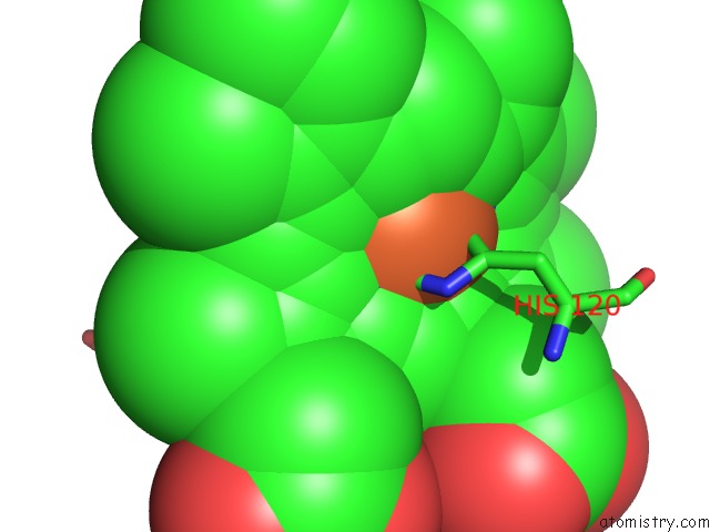
Mono view
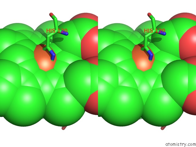
Stereo pair view
Mono view
Stereo pair view
A full contact list of Iron with other atoms in the Fe binding
site number 1 of 3D Structure of Ferric Methanosarcina Acetivorans Protoglobin Y61A Mutant with Unknown Ligand within 5.0Å range:
|
Reference:
A.Pesce,
L.Tilleman,
S.Dewilde,
P.Ascenzi,
M.Coletta,
C.Ciaccio,
S.Bruno,
L.Moens,
M.Bolognesi,
M.Nardini.
Structural Heterogeneity and Ligand Gating in Ferric Methanosarcina Acetivorans Protoglobin Mutants. Iubmb Life V. 63 287 2011.
ISSN: ISSN 1521-6543
PubMed: 21618401
DOI: 10.1002/IUB.484
Page generated: Sun Aug 4 19:00:12 2024
ISSN: ISSN 1521-6543
PubMed: 21618401
DOI: 10.1002/IUB.484
Last articles
Zn in 9MJ5Zn in 9HNW
Zn in 9G0L
Zn in 9FNE
Zn in 9DZN
Zn in 9E0I
Zn in 9D32
Zn in 9DAK
Zn in 8ZXC
Zn in 8ZUF