Iron »
PDB 3r1a-3rmk »
3r60 »
Iron in PDB 3r60: Structure of the Mntr FE2+ Complex
Protein crystallography data
The structure of Structure of the Mntr FE2+ Complex, PDB code: 3r60
was solved by
A.Glasfeld,
M.B.Brophy,
J.I.Kliegman,
S.L.Griner,
J.C.Nix,
with X-Ray Crystallography technique. A brief refinement statistics is given in the table below:
| Resolution Low / High (Å) | 21.82 / 1.80 |
| Space group | P 1 21 1 |
| Cell size a, b, c (Å), α, β, γ (°) | 49.680, 45.900, 74.690, 90.00, 93.50, 90.00 |
| R / Rfree (%) | 23.9 / 28.5 |
Iron Binding Sites:
The binding sites of Iron atom in the Structure of the Mntr FE2+ Complex
(pdb code 3r60). This binding sites where shown within
5.0 Angstroms radius around Iron atom.
In total 2 binding sites of Iron where determined in the Structure of the Mntr FE2+ Complex, PDB code: 3r60:
Jump to Iron binding site number: 1; 2;
In total 2 binding sites of Iron where determined in the Structure of the Mntr FE2+ Complex, PDB code: 3r60:
Jump to Iron binding site number: 1; 2;
Iron binding site 1 out of 2 in 3r60
Go back to
Iron binding site 1 out
of 2 in the Structure of the Mntr FE2+ Complex
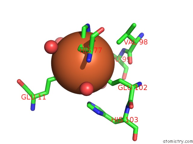
Mono view
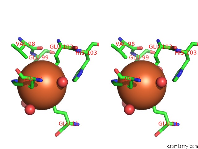
Stereo pair view
Mono view
Stereo pair view
A full contact list of Iron with other atoms in the Fe binding
site number 1 of Structure of the Mntr FE2+ Complex within 5.0Å range:
|
Iron binding site 2 out of 2 in 3r60
Go back to
Iron binding site 2 out
of 2 in the Structure of the Mntr FE2+ Complex
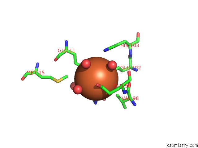
Mono view
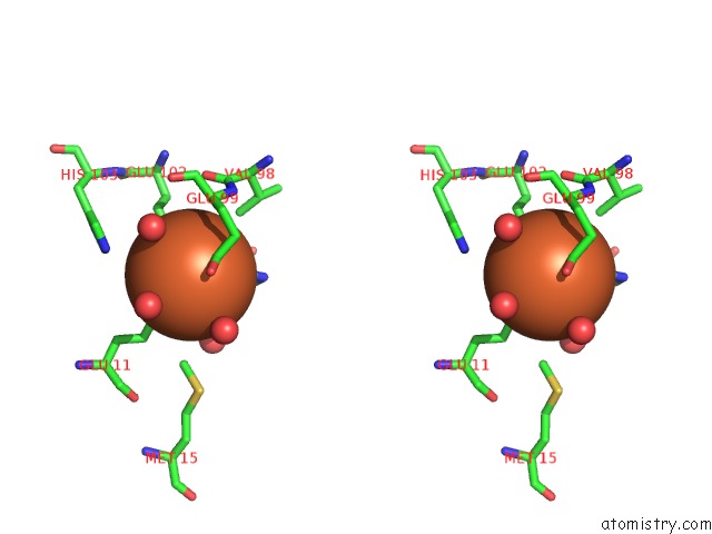
Stereo pair view
Mono view
Stereo pair view
A full contact list of Iron with other atoms in the Fe binding
site number 2 of Structure of the Mntr FE2+ Complex within 5.0Å range:
|
Reference:
A.M.Mcguire,
B.J.Cuthbert,
Z.Ma,
K.D.Grauer-Gray,
M.Brunjes Brophy,
K.A.Spear,
S.Soonsanga,
J.I.Kliegman,
S.L.Griner,
J.D.Helmann,
A.Glasfeld.
Roles of the A and C Sites in the Manganese-Specific Activation of Mntr. Biochemistry V. 52 701 2013.
ISSN: ISSN 0006-2960
PubMed: 23298157
DOI: 10.1021/BI301550T
Page generated: Sun Aug 4 19:11:20 2024
ISSN: ISSN 0006-2960
PubMed: 23298157
DOI: 10.1021/BI301550T
Last articles
Zn in 9J0NZn in 9J0O
Zn in 9J0P
Zn in 9FJX
Zn in 9EKB
Zn in 9C0F
Zn in 9CAH
Zn in 9CH0
Zn in 9CH3
Zn in 9CH1