Iron »
PDB 3r1a-3rmk »
3rfa »
Iron in PDB 3rfa: X-Ray Structure of Rlmn From Escherichia Coli in Complex with S- Adenosylmethionine
Enzymatic activity of X-Ray Structure of Rlmn From Escherichia Coli in Complex with S- Adenosylmethionine
All present enzymatic activity of X-Ray Structure of Rlmn From Escherichia Coli in Complex with S- Adenosylmethionine:
2.1.1.192;
2.1.1.192;
Protein crystallography data
The structure of X-Ray Structure of Rlmn From Escherichia Coli in Complex with S- Adenosylmethionine, PDB code: 3rfa
was solved by
A.K.Boal,
T.L.Grove,
M.I.Mclaughlin,
N.Yennawar,
S.J.Booker,
A.C.Rosenzweig,
with X-Ray Crystallography technique. A brief refinement statistics is given in the table below:
| Resolution Low / High (Å) | 30.00 / 2.05 |
| Space group | P 21 21 21 |
| Cell size a, b, c (Å), α, β, γ (°) | 55.180, 55.621, 252.179, 90.00, 90.00, 90.00 |
| R / Rfree (%) | 20.2 / 24.1 |
Iron Binding Sites:
The binding sites of Iron atom in the X-Ray Structure of Rlmn From Escherichia Coli in Complex with S- Adenosylmethionine
(pdb code 3rfa). This binding sites where shown within
5.0 Angstroms radius around Iron atom.
In total 8 binding sites of Iron where determined in the X-Ray Structure of Rlmn From Escherichia Coli in Complex with S- Adenosylmethionine, PDB code: 3rfa:
Jump to Iron binding site number: 1; 2; 3; 4; 5; 6; 7; 8;
In total 8 binding sites of Iron where determined in the X-Ray Structure of Rlmn From Escherichia Coli in Complex with S- Adenosylmethionine, PDB code: 3rfa:
Jump to Iron binding site number: 1; 2; 3; 4; 5; 6; 7; 8;
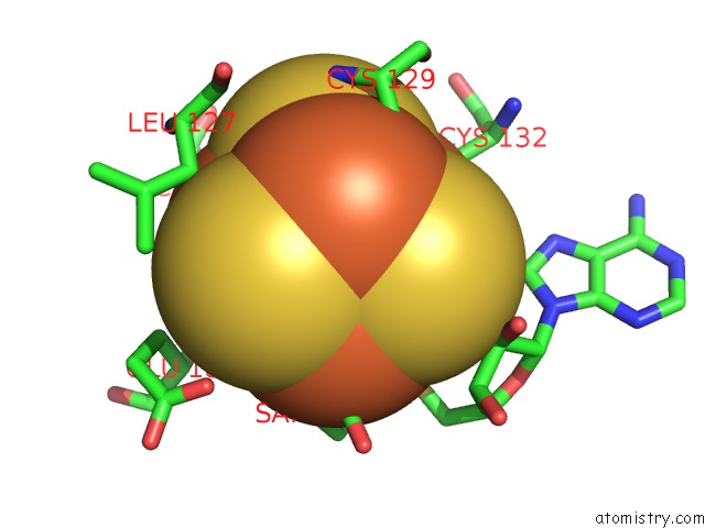
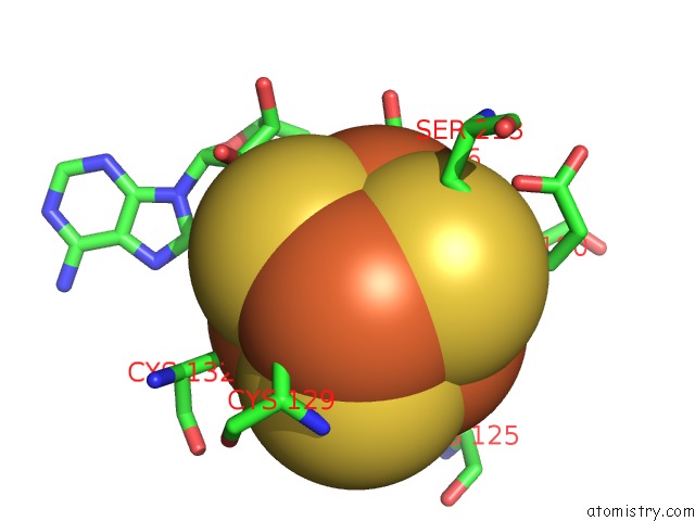
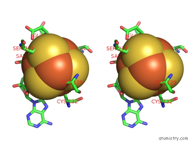
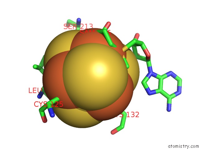
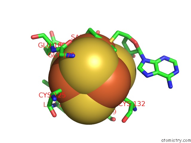
Iron binding site 1 out of 8 in 3rfa
Go back to
Iron binding site 1 out
of 8 in the X-Ray Structure of Rlmn From Escherichia Coli in Complex with S- Adenosylmethionine
Mono view
Stereo pair view
Mono view
Stereo pair view
A full contact list of Iron with other atoms in the Fe binding
site number 1 of X-Ray Structure of Rlmn From Escherichia Coli in Complex with S- Adenosylmethionine within 5.0Å range:
|
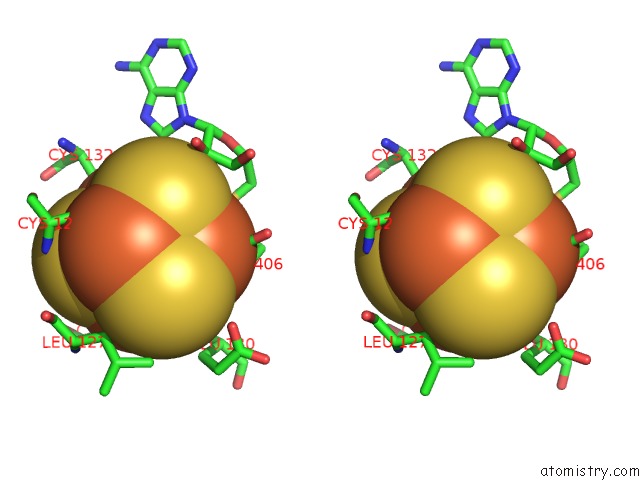
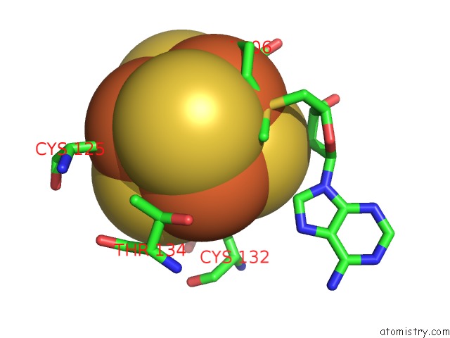
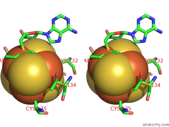
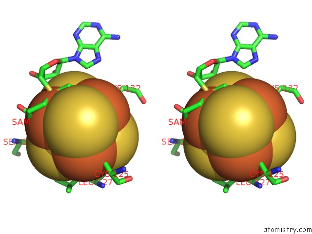
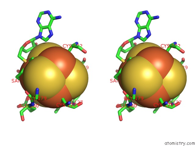
Iron binding site 2 out of 8 in 3rfa
Go back to
Iron binding site 2 out
of 8 in the X-Ray Structure of Rlmn From Escherichia Coli in Complex with S- Adenosylmethionine
Mono view
Stereo pair view
Mono view
Stereo pair view
A full contact list of Iron with other atoms in the Fe binding
site number 2 of X-Ray Structure of Rlmn From Escherichia Coli in Complex with S- Adenosylmethionine within 5.0Å range:
|
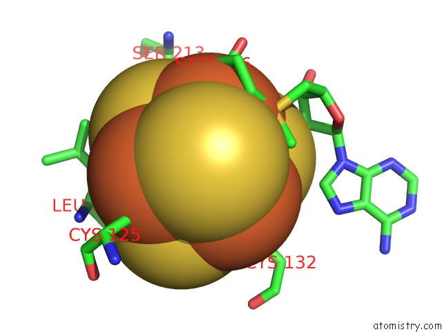
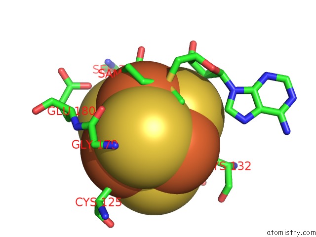
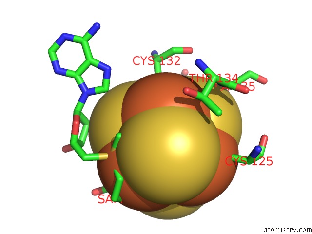
Iron binding site 3 out of 8 in 3rfa
Go back to
Iron binding site 3 out
of 8 in the X-Ray Structure of Rlmn From Escherichia Coli in Complex with S- Adenosylmethionine
Mono view
Stereo pair view
Mono view
Stereo pair view
A full contact list of Iron with other atoms in the Fe binding
site number 3 of X-Ray Structure of Rlmn From Escherichia Coli in Complex with S- Adenosylmethionine within 5.0Å range:
|
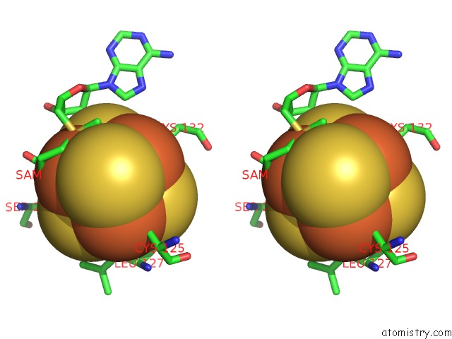
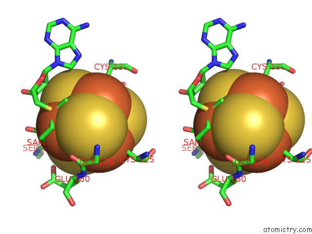
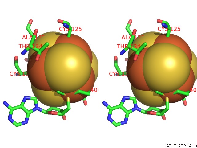
Iron binding site 4 out of 8 in 3rfa
Go back to
Iron binding site 4 out
of 8 in the X-Ray Structure of Rlmn From Escherichia Coli in Complex with S- Adenosylmethionine
Mono view
Stereo pair view
Mono view
Stereo pair view
A full contact list of Iron with other atoms in the Fe binding
site number 4 of X-Ray Structure of Rlmn From Escherichia Coli in Complex with S- Adenosylmethionine within 5.0Å range:
|
Iron binding site 5 out of 8 in 3rfa
Go back to
Iron binding site 5 out
of 8 in the X-Ray Structure of Rlmn From Escherichia Coli in Complex with S- Adenosylmethionine
Mono view
Stereo pair view
Mono view
Stereo pair view
A full contact list of Iron with other atoms in the Fe binding
site number 5 of X-Ray Structure of Rlmn From Escherichia Coli in Complex with S- Adenosylmethionine within 5.0Å range:
|
Iron binding site 6 out of 8 in 3rfa
Go back to
Iron binding site 6 out
of 8 in the X-Ray Structure of Rlmn From Escherichia Coli in Complex with S- Adenosylmethionine
Mono view
Stereo pair view
Mono view
Stereo pair view
A full contact list of Iron with other atoms in the Fe binding
site number 6 of X-Ray Structure of Rlmn From Escherichia Coli in Complex with S- Adenosylmethionine within 5.0Å range:
|
Iron binding site 7 out of 8 in 3rfa
Go back to
Iron binding site 7 out
of 8 in the X-Ray Structure of Rlmn From Escherichia Coli in Complex with S- Adenosylmethionine
Mono view
Stereo pair view
Mono view
Stereo pair view
A full contact list of Iron with other atoms in the Fe binding
site number 7 of X-Ray Structure of Rlmn From Escherichia Coli in Complex with S- Adenosylmethionine within 5.0Å range:
|
Iron binding site 8 out of 8 in 3rfa
Go back to
Iron binding site 8 out
of 8 in the X-Ray Structure of Rlmn From Escherichia Coli in Complex with S- Adenosylmethionine
Mono view
Stereo pair view
Mono view
Stereo pair view
A full contact list of Iron with other atoms in the Fe binding
site number 8 of X-Ray Structure of Rlmn From Escherichia Coli in Complex with S- Adenosylmethionine within 5.0Å range:
|
Reference:
A.K.Boal,
T.L.Grove,
M.I.Mclaughlin,
N.H.Yennawar,
S.J.Booker,
A.C.Rosenzweig.
Structural Basis For Methyl Transfer By A Radical Sam Enzyme. Science V. 332 1089 2011.
ISSN: ISSN 0036-8075
PubMed: 21527678
DOI: 10.1126/SCIENCE.1205358
Page generated: Sun Aug 4 19:13:37 2024
ISSN: ISSN 0036-8075
PubMed: 21527678
DOI: 10.1126/SCIENCE.1205358
Last articles
Zn in 9JYWZn in 9IR4
Zn in 9IR3
Zn in 9GMX
Zn in 9GMW
Zn in 9JEJ
Zn in 9ERF
Zn in 9ERE
Zn in 9EGV
Zn in 9EGW