Iron »
PDB 3r1a-3rmk »
3riw »
Iron in PDB 3riw: The Crystal Structure of Leishmania Major Peroxidase Mutant C197T
Enzymatic activity of The Crystal Structure of Leishmania Major Peroxidase Mutant C197T
All present enzymatic activity of The Crystal Structure of Leishmania Major Peroxidase Mutant C197T:
1.11.1.11;
1.11.1.11;
Protein crystallography data
The structure of The Crystal Structure of Leishmania Major Peroxidase Mutant C197T, PDB code: 3riw
was solved by
V.S.Jasion,
H.Li,
T.L.Poulos,
with X-Ray Crystallography technique. A brief refinement statistics is given in the table below:
| Resolution Low / High (Å) | 35.43 / 2.37 |
| Space group | P 21 21 21 |
| Cell size a, b, c (Å), α, β, γ (°) | 45.819, 77.766, 160.580, 90.00, 90.00, 90.00 |
| R / Rfree (%) | 17.8 / 24.1 |
Other elements in 3riw:
The structure of The Crystal Structure of Leishmania Major Peroxidase Mutant C197T also contains other interesting chemical elements:
| Potassium | (K) | 2 atoms |
| Calcium | (Ca) | 2 atoms |
Iron Binding Sites:
The binding sites of Iron atom in the The Crystal Structure of Leishmania Major Peroxidase Mutant C197T
(pdb code 3riw). This binding sites where shown within
5.0 Angstroms radius around Iron atom.
In total 2 binding sites of Iron where determined in the The Crystal Structure of Leishmania Major Peroxidase Mutant C197T, PDB code: 3riw:
Jump to Iron binding site number: 1; 2;
In total 2 binding sites of Iron where determined in the The Crystal Structure of Leishmania Major Peroxidase Mutant C197T, PDB code: 3riw:
Jump to Iron binding site number: 1; 2;
Iron binding site 1 out of 2 in 3riw
Go back to
Iron binding site 1 out
of 2 in the The Crystal Structure of Leishmania Major Peroxidase Mutant C197T
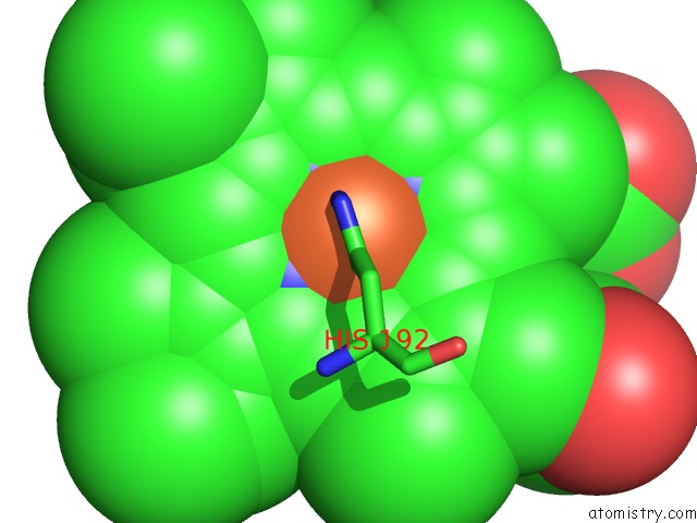
Mono view
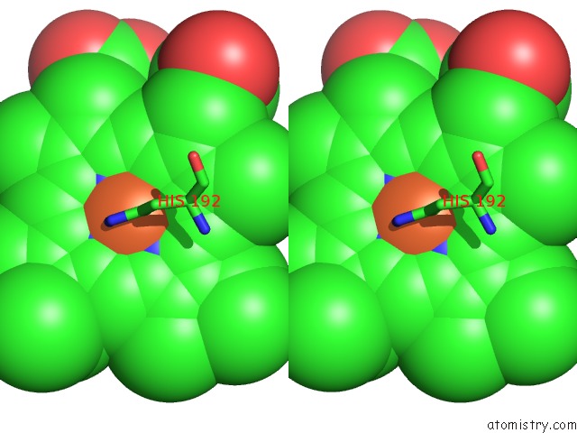
Stereo pair view
Mono view
Stereo pair view
A full contact list of Iron with other atoms in the Fe binding
site number 1 of The Crystal Structure of Leishmania Major Peroxidase Mutant C197T within 5.0Å range:
|
Iron binding site 2 out of 2 in 3riw
Go back to
Iron binding site 2 out
of 2 in the The Crystal Structure of Leishmania Major Peroxidase Mutant C197T
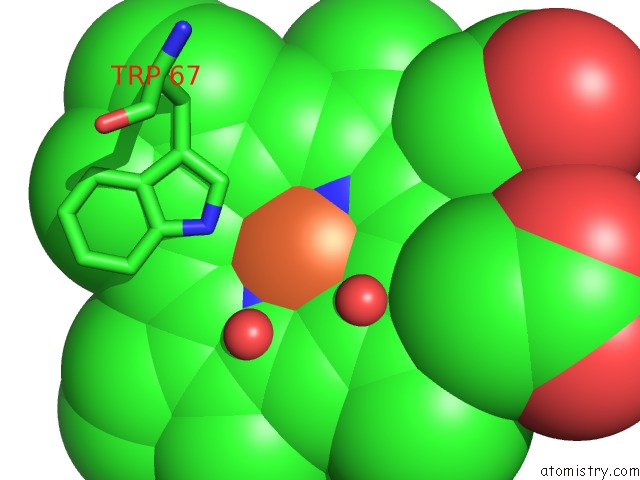
Mono view
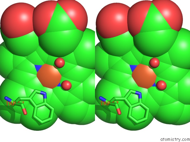
Stereo pair view
Mono view
Stereo pair view
A full contact list of Iron with other atoms in the Fe binding
site number 2 of The Crystal Structure of Leishmania Major Peroxidase Mutant C197T within 5.0Å range:
|
Reference:
V.S.Jasion,
J.A.Polanco,
Y.T.Meharenna,
H.Li,
T.L.Poulos.
Crystal Structure of Leishmania Major Peroxidase and Characterization of the Compound I Tryptophan Radical. J.Biol.Chem. V. 286 24608 2011.
ISSN: ISSN 0021-9258
PubMed: 21566139
DOI: 10.1074/JBC.M111.230524
Page generated: Sun Aug 4 19:19:09 2024
ISSN: ISSN 0021-9258
PubMed: 21566139
DOI: 10.1074/JBC.M111.230524
Last articles
Fe in 2YXOFe in 2YRS
Fe in 2YXC
Fe in 2YNM
Fe in 2YVJ
Fe in 2YP1
Fe in 2YU2
Fe in 2YU1
Fe in 2YQB
Fe in 2YOO