Iron »
PDB 3rmz-3s66 »
3rnb »
Iron in PDB 3rnb: Structure of the Toluene/O-Xylene Monooxygenase Hydroxylase T201S/F176W Double Mutant
Protein crystallography data
The structure of Structure of the Toluene/O-Xylene Monooxygenase Hydroxylase T201S/F176W Double Mutant, PDB code: 3rnb
was solved by
G.Gucinski,
W.J.Song,
S.J.Lippard,
M.H.Sazinsky,
with X-Ray Crystallography technique. A brief refinement statistics is given in the table below:
| Resolution Low / High (Å) | 44.99 / 2.64 |
| Space group | P 31 2 1 |
| Cell size a, b, c (Å), α, β, γ (°) | 183.162, 183.162, 68.081, 90.00, 90.00, 120.00 |
| R / Rfree (%) | 17.5 / 23.5 |
Iron Binding Sites:
The binding sites of Iron atom in the Structure of the Toluene/O-Xylene Monooxygenase Hydroxylase T201S/F176W Double Mutant
(pdb code 3rnb). This binding sites where shown within
5.0 Angstroms radius around Iron atom.
In total 2 binding sites of Iron where determined in the Structure of the Toluene/O-Xylene Monooxygenase Hydroxylase T201S/F176W Double Mutant, PDB code: 3rnb:
Jump to Iron binding site number: 1; 2;
In total 2 binding sites of Iron where determined in the Structure of the Toluene/O-Xylene Monooxygenase Hydroxylase T201S/F176W Double Mutant, PDB code: 3rnb:
Jump to Iron binding site number: 1; 2;
Iron binding site 1 out of 2 in 3rnb
Go back to
Iron binding site 1 out
of 2 in the Structure of the Toluene/O-Xylene Monooxygenase Hydroxylase T201S/F176W Double Mutant
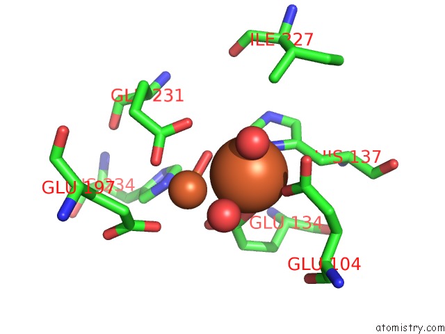
Mono view
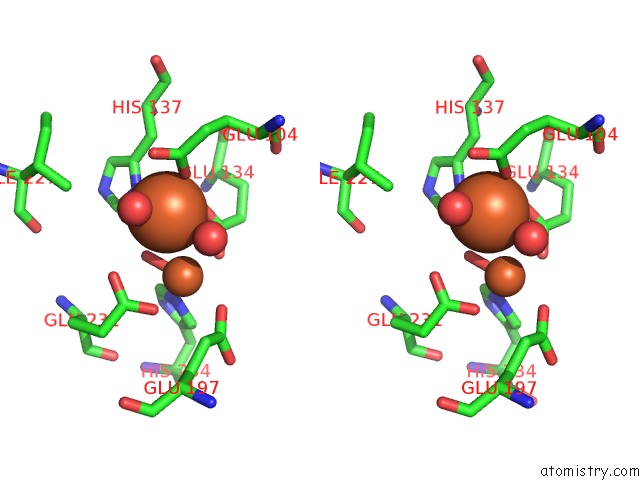
Stereo pair view
Mono view
Stereo pair view
A full contact list of Iron with other atoms in the Fe binding
site number 1 of Structure of the Toluene/O-Xylene Monooxygenase Hydroxylase T201S/F176W Double Mutant within 5.0Å range:
|
Iron binding site 2 out of 2 in 3rnb
Go back to
Iron binding site 2 out
of 2 in the Structure of the Toluene/O-Xylene Monooxygenase Hydroxylase T201S/F176W Double Mutant
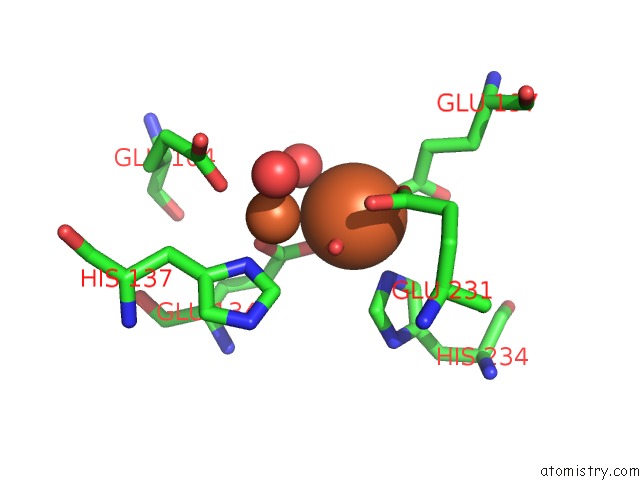
Mono view

Stereo pair view
Mono view
Stereo pair view
A full contact list of Iron with other atoms in the Fe binding
site number 2 of Structure of the Toluene/O-Xylene Monooxygenase Hydroxylase T201S/F176W Double Mutant within 5.0Å range:
|
Reference:
W.J.Song,
G.Gucinski,
M.H.Sazinsky,
S.J.Lippard.
Tracking A Defined Route For O2 Migration in A Dioxygen-Activating Diiron Enzyme. Proc.Natl.Acad.Sci.Usa V. 108 14795 2011.
ISSN: ISSN 0027-8424
PubMed: 21859951
DOI: 10.1073/PNAS.1106514108
Page generated: Sun Aug 4 19:41:45 2024
ISSN: ISSN 0027-8424
PubMed: 21859951
DOI: 10.1073/PNAS.1106514108
Last articles
Zn in 9J0NZn in 9J0O
Zn in 9J0P
Zn in 9FJX
Zn in 9EKB
Zn in 9C0F
Zn in 9CAH
Zn in 9CH0
Zn in 9CH3
Zn in 9CH1