Iron »
PDB 3rmz-3s66 »
3rwl »
Iron in PDB 3rwl: Structure of P450PYR Hydroxylase
Enzymatic activity of Structure of P450PYR Hydroxylase
All present enzymatic activity of Structure of P450PYR Hydroxylase:
1.14.15.3;
1.14.15.3;
Protein crystallography data
The structure of Structure of P450PYR Hydroxylase, PDB code: 3rwl
was solved by
G.Pompidor,
with X-Ray Crystallography technique. A brief refinement statistics is given in the table below:
| Resolution Low / High (Å) | 48.76 / 2.00 |
| Space group | P 31 |
| Cell size a, b, c (Å), α, β, γ (°) | 112.604, 112.604, 41.964, 90.00, 90.00, 120.00 |
| R / Rfree (%) | 16 / 19.6 |
Iron Binding Sites:
The binding sites of Iron atom in the Structure of P450PYR Hydroxylase
(pdb code 3rwl). This binding sites where shown within
5.0 Angstroms radius around Iron atom.
In total only one binding site of Iron was determined in the Structure of P450PYR Hydroxylase, PDB code: 3rwl:
In total only one binding site of Iron was determined in the Structure of P450PYR Hydroxylase, PDB code: 3rwl:
Iron binding site 1 out of 1 in 3rwl
Go back to
Iron binding site 1 out
of 1 in the Structure of P450PYR Hydroxylase
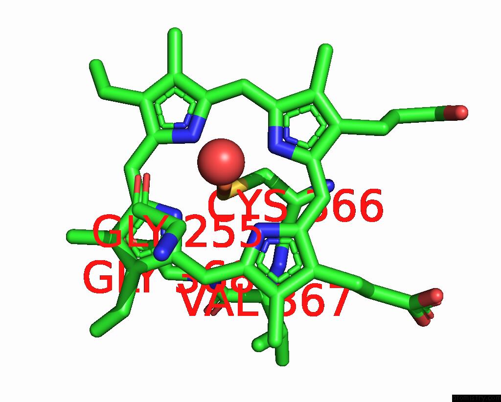
Mono view
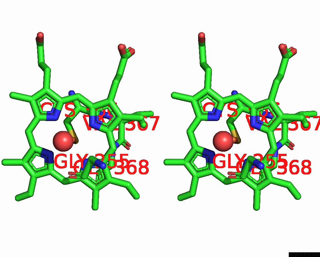
Stereo pair view
Mono view
Stereo pair view
A full contact list of Iron with other atoms in the Fe binding
site number 1 of Structure of P450PYR Hydroxylase within 5.0Å range:
|
Reference:
S.Q.Pham,
G.Pompidor,
J.Liu,
X.D.Li,
Z.Li.
Evolving P450PYR Hydroxylase For Highly Enantioselective Hydroxylation at Non-Activated Carbon Atom. Chem.Commun.(Camb.) V. 48 4618 2012.
ISSN: ISSN 1359-7345
PubMed: 22430002
DOI: 10.1039/C2CC30779K
Page generated: Sun Aug 4 19:46:03 2024
ISSN: ISSN 1359-7345
PubMed: 22430002
DOI: 10.1039/C2CC30779K
Last articles
Zn in 9J0NZn in 9J0O
Zn in 9J0P
Zn in 9FJX
Zn in 9EKB
Zn in 9C0F
Zn in 9CAH
Zn in 9CH0
Zn in 9CH3
Zn in 9CH1