Iron »
PDB 3rmz-3s66 »
3s65 »
Iron in PDB 3s65: Structures and Oxygen Affinities of Crystalline Human Hemoglobin C (BETA6 Lys) in the R2 Quaternary Structures
Protein crystallography data
The structure of Structures and Oxygen Affinities of Crystalline Human Hemoglobin C (BETA6 Lys) in the R2 Quaternary Structures, PDB code: 3s65
was solved by
N.Shibayama,
K.Sugiyama,
S.Y.Park,
with X-Ray Crystallography technique. A brief refinement statistics is given in the table below:
| Resolution Low / High (Å) | 19.40 / 1.80 |
| Space group | P 21 21 21 |
| Cell size a, b, c (Å), α, β, γ (°) | 57.784, 58.748, 172.869, 90.00, 90.00, 90.00 |
| R / Rfree (%) | 25 / 30.5 |
Iron Binding Sites:
The binding sites of Iron atom in the Structures and Oxygen Affinities of Crystalline Human Hemoglobin C (BETA6 Lys) in the R2 Quaternary Structures
(pdb code 3s65). This binding sites where shown within
5.0 Angstroms radius around Iron atom.
In total 4 binding sites of Iron where determined in the Structures and Oxygen Affinities of Crystalline Human Hemoglobin C (BETA6 Lys) in the R2 Quaternary Structures, PDB code: 3s65:
Jump to Iron binding site number: 1; 2; 3; 4;
In total 4 binding sites of Iron where determined in the Structures and Oxygen Affinities of Crystalline Human Hemoglobin C (BETA6 Lys) in the R2 Quaternary Structures, PDB code: 3s65:
Jump to Iron binding site number: 1; 2; 3; 4;
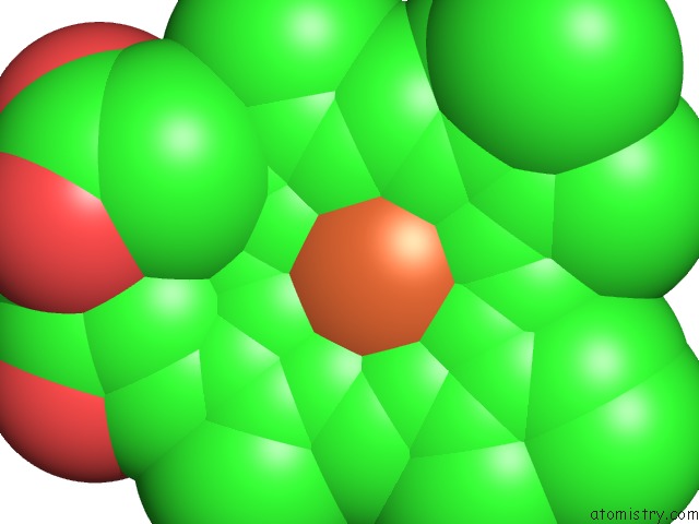
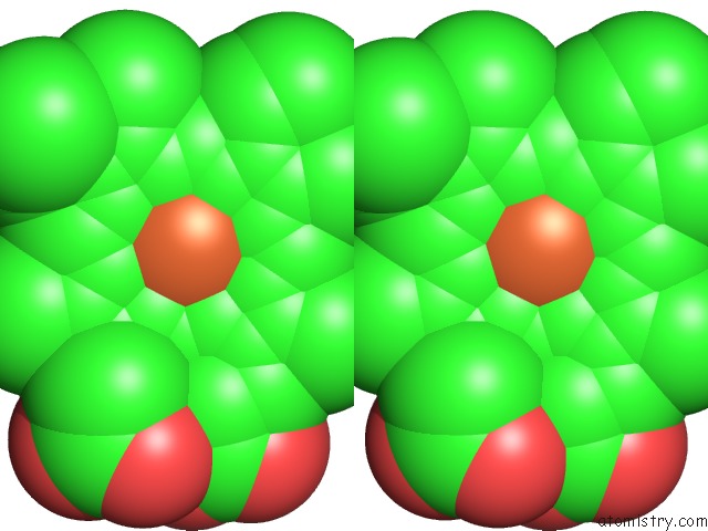
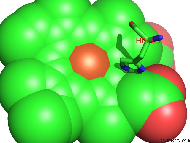
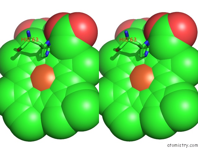
Iron binding site 1 out of 4 in 3s65
Go back to
Iron binding site 1 out
of 4 in the Structures and Oxygen Affinities of Crystalline Human Hemoglobin C (BETA6 Lys) in the R2 Quaternary Structures
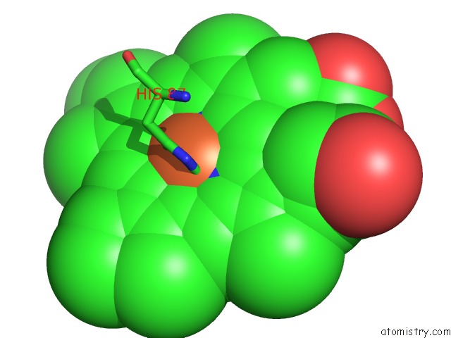
Mono view
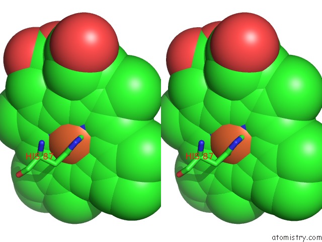
Stereo pair view
Mono view
Stereo pair view
A full contact list of Iron with other atoms in the Fe binding
site number 1 of Structures and Oxygen Affinities of Crystalline Human Hemoglobin C (BETA6 Lys) in the R2 Quaternary Structures within 5.0Å range:
|
Iron binding site 2 out of 4 in 3s65
Go back to
Iron binding site 2 out
of 4 in the Structures and Oxygen Affinities of Crystalline Human Hemoglobin C (BETA6 Lys) in the R2 Quaternary Structures
Mono view
Stereo pair view
Mono view
Stereo pair view
A full contact list of Iron with other atoms in the Fe binding
site number 2 of Structures and Oxygen Affinities of Crystalline Human Hemoglobin C (BETA6 Lys) in the R2 Quaternary Structures within 5.0Å range:
|
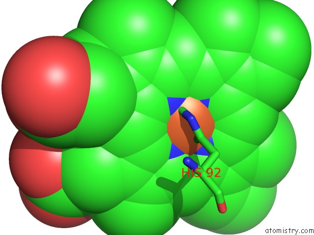
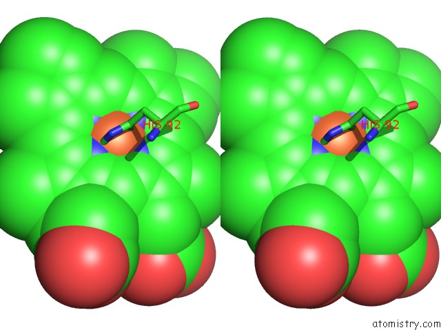
Iron binding site 3 out of 4 in 3s65
Go back to
Iron binding site 3 out
of 4 in the Structures and Oxygen Affinities of Crystalline Human Hemoglobin C (BETA6 Lys) in the R2 Quaternary Structures
Mono view
Stereo pair view
Mono view
Stereo pair view
A full contact list of Iron with other atoms in the Fe binding
site number 3 of Structures and Oxygen Affinities of Crystalline Human Hemoglobin C (BETA6 Lys) in the R2 Quaternary Structures within 5.0Å range:
|
Iron binding site 4 out of 4 in 3s65
Go back to
Iron binding site 4 out
of 4 in the Structures and Oxygen Affinities of Crystalline Human Hemoglobin C (BETA6 Lys) in the R2 Quaternary Structures
Mono view
Stereo pair view
Mono view
Stereo pair view
A full contact list of Iron with other atoms in the Fe binding
site number 4 of Structures and Oxygen Affinities of Crystalline Human Hemoglobin C (BETA6 Lys) in the R2 Quaternary Structures within 5.0Å range:
|
Reference:
N.Shibayama,
K.Sugiyama,
S.Y.Park.
Structures and Oxygen Affinities of Crystalline Human Hemoglobin C (BETA6 Glu->Lys) in the R and R2 Quaternary Structure To Be Published.
Page generated: Sun Aug 4 19:50:08 2024
Last articles
F in 4OZ3F in 4P45
F in 4P1U
F in 4P44
F in 4P2H
F in 4OWO
F in 4OZ2
F in 4OYT
F in 4OWN
F in 4OYS