Iron »
PDB 3s6b-3sxt »
3sel »
Iron in PDB 3sel: Ppca M58N Mutant
Protein crystallography data
The structure of Ppca M58N Mutant, PDB code: 3sel
was solved by
P.R.Pokkuluri,
M.Schiffer,
with X-Ray Crystallography technique. A brief refinement statistics is given in the table below:
| Resolution Low / High (Å) | 30.00 / 2.10 |
| Space group | P 43 2 2 |
| Cell size a, b, c (Å), α, β, γ (°) | 32.420, 32.420, 177.450, 90.00, 90.00, 90.00 |
| R / Rfree (%) | 17.6 / 22.3 |
Iron Binding Sites:
The binding sites of Iron atom in the Ppca M58N Mutant
(pdb code 3sel). This binding sites where shown within
5.0 Angstroms radius around Iron atom.
In total 3 binding sites of Iron where determined in the Ppca M58N Mutant, PDB code: 3sel:
Jump to Iron binding site number: 1; 2; 3;
In total 3 binding sites of Iron where determined in the Ppca M58N Mutant, PDB code: 3sel:
Jump to Iron binding site number: 1; 2; 3;
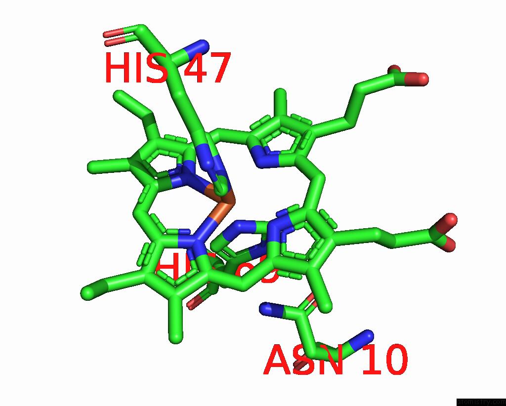
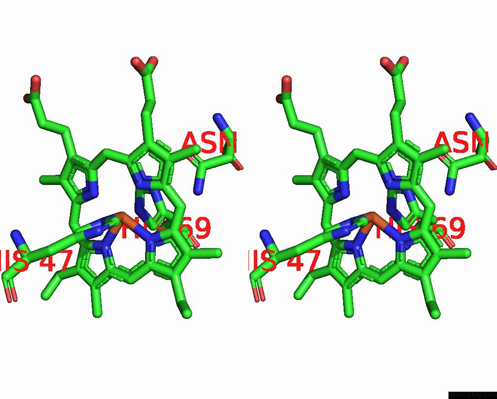
Iron binding site 1 out of 3 in 3sel
Go back to
Iron binding site 1 out
of 3 in the Ppca M58N Mutant
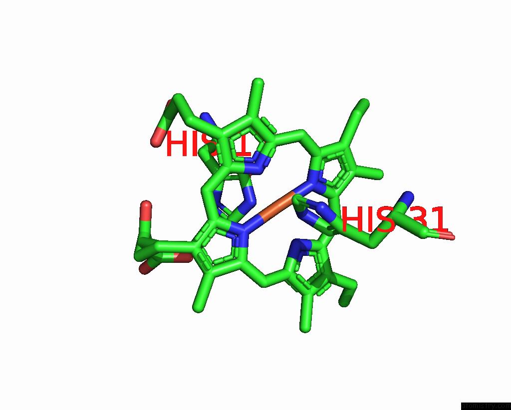
Mono view
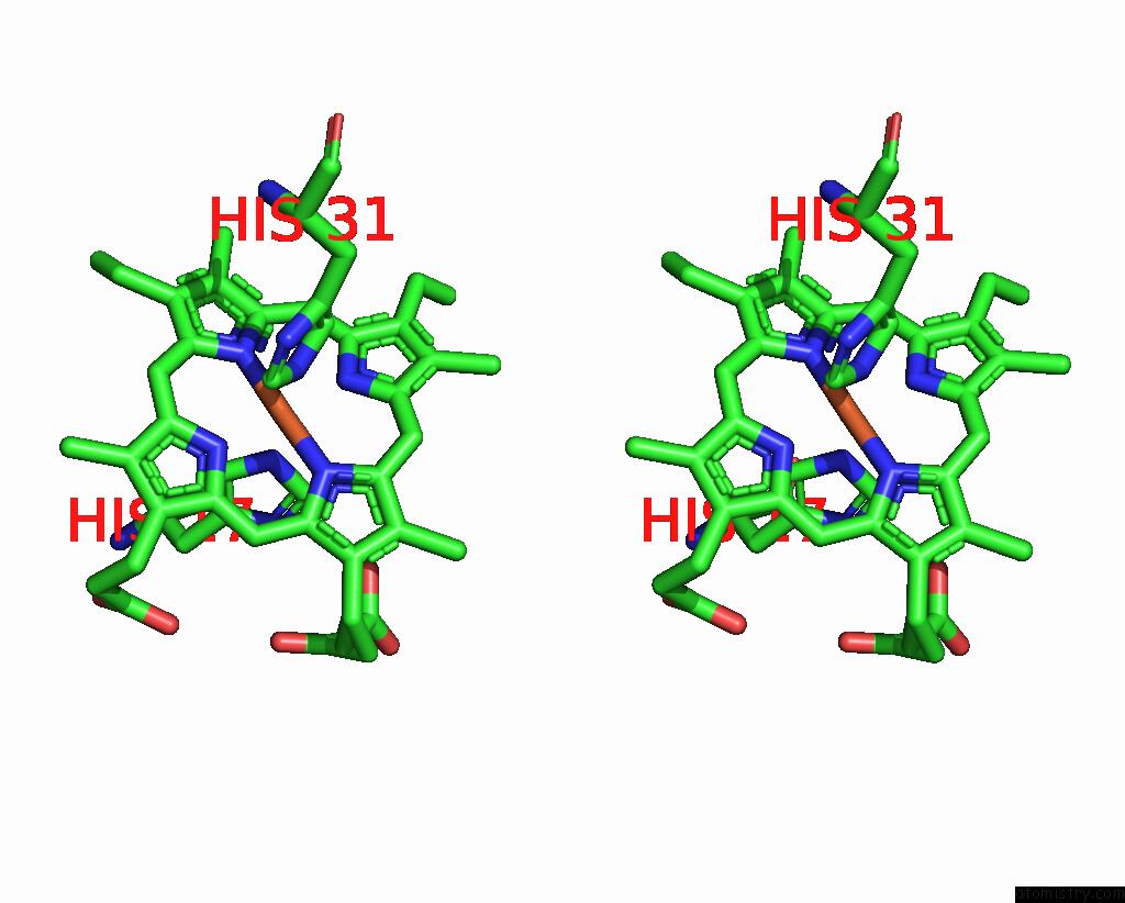
Stereo pair view
Mono view
Stereo pair view
A full contact list of Iron with other atoms in the Fe binding
site number 1 of Ppca M58N Mutant within 5.0Å range:
|
Iron binding site 2 out of 3 in 3sel
Go back to
Iron binding site 2 out
of 3 in the Ppca M58N Mutant
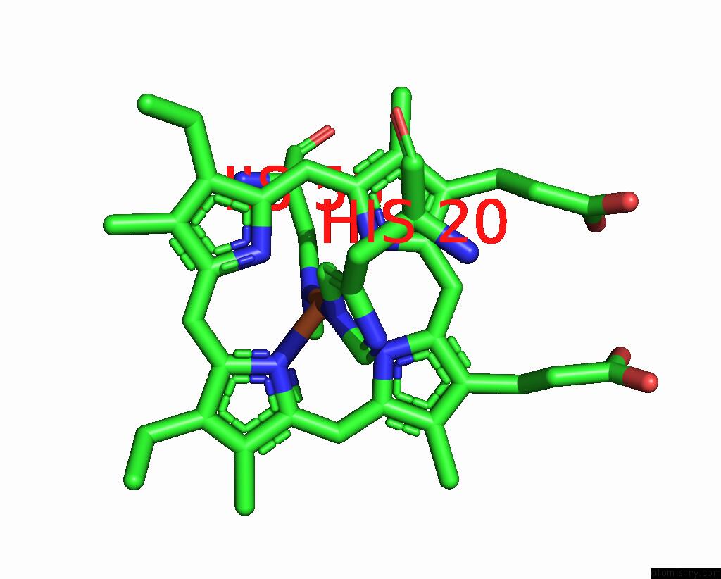
Mono view
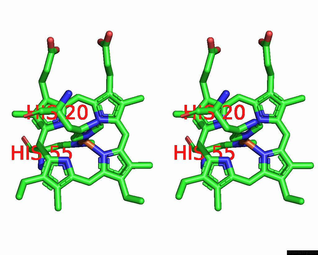
Stereo pair view
Mono view
Stereo pair view
A full contact list of Iron with other atoms in the Fe binding
site number 2 of Ppca M58N Mutant within 5.0Å range:
|
Iron binding site 3 out of 3 in 3sel
Go back to
Iron binding site 3 out
of 3 in the Ppca M58N Mutant
Mono view
Stereo pair view
Mono view
Stereo pair view
A full contact list of Iron with other atoms in the Fe binding
site number 3 of Ppca M58N Mutant within 5.0Å range:
|
Reference:
P.R.Pokkuluri,
X.Yang,
Y.Y.Londer,
M.Schiffer.
Pitfalls in the Interpretation of Structural Changes in Mutant Proteins From Crystal Structures. J.Struct.Funct.Genom. V. 13 227 2012.
ISSN: ISSN 1345-711X
PubMed: 23099666
DOI: 10.1007/S10969-012-9147-1
Page generated: Sun Aug 4 19:55:05 2024
ISSN: ISSN 1345-711X
PubMed: 23099666
DOI: 10.1007/S10969-012-9147-1
Last articles
Zn in 9J0NZn in 9J0O
Zn in 9J0P
Zn in 9FJX
Zn in 9EKB
Zn in 9C0F
Zn in 9CAH
Zn in 9CH0
Zn in 9CH3
Zn in 9CH1