Iron »
PDB 3s6b-3sxt »
3sj5 »
Iron in PDB 3sj5: I5F Mutant Structure of T. Tengcongensis H-Nox
Protein crystallography data
The structure of I5F Mutant Structure of T. Tengcongensis H-Nox, PDB code: 3sj5
was solved by
E.E.Weinert,
C.M.Phillips-Piro,
R.Tran,
R.A.Mathies,
M.A.Marletta,
with X-Ray Crystallography technique. A brief refinement statistics is given in the table below:
| Resolution Low / High (Å) | 47.20 / 1.67 |
| Space group | P 1 21 1 |
| Cell size a, b, c (Å), α, β, γ (°) | 44.585, 67.116, 66.428, 90.00, 92.01, 90.00 |
| R / Rfree (%) | 19.7 / 24.7 |
Iron Binding Sites:
The binding sites of Iron atom in the I5F Mutant Structure of T. Tengcongensis H-Nox
(pdb code 3sj5). This binding sites where shown within
5.0 Angstroms radius around Iron atom.
In total 2 binding sites of Iron where determined in the I5F Mutant Structure of T. Tengcongensis H-Nox, PDB code: 3sj5:
Jump to Iron binding site number: 1; 2;
In total 2 binding sites of Iron where determined in the I5F Mutant Structure of T. Tengcongensis H-Nox, PDB code: 3sj5:
Jump to Iron binding site number: 1; 2;
Iron binding site 1 out of 2 in 3sj5
Go back to
Iron binding site 1 out
of 2 in the I5F Mutant Structure of T. Tengcongensis H-Nox
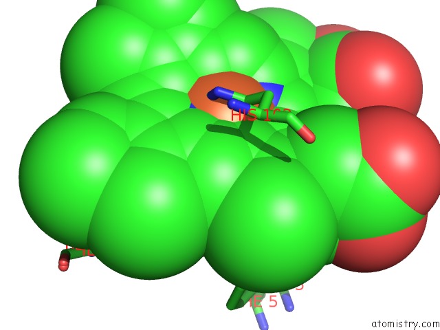
Mono view
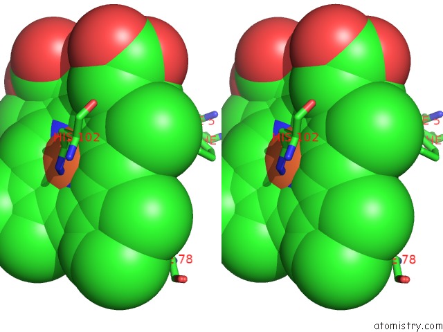
Stereo pair view
Mono view
Stereo pair view
A full contact list of Iron with other atoms in the Fe binding
site number 1 of I5F Mutant Structure of T. Tengcongensis H-Nox within 5.0Å range:
|
Iron binding site 2 out of 2 in 3sj5
Go back to
Iron binding site 2 out
of 2 in the I5F Mutant Structure of T. Tengcongensis H-Nox
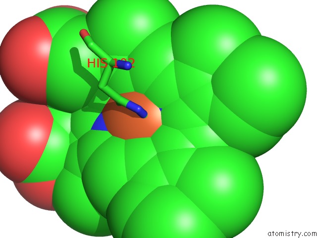
Mono view
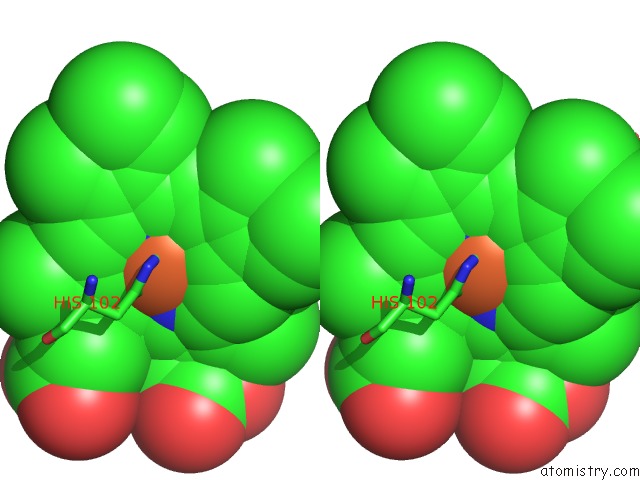
Stereo pair view
Mono view
Stereo pair view
A full contact list of Iron with other atoms in the Fe binding
site number 2 of I5F Mutant Structure of T. Tengcongensis H-Nox within 5.0Å range:
|
Reference:
E.E.Weinert,
C.M.Phillips-Piro,
R.Tran,
R.A.Mathies,
M.A.Marletta.
Controlling Conformational Flexibility of An O2-Binding H-Nox Domain. Biochemistry V. 50 6832 2011.
ISSN: ISSN 0006-2960
PubMed: 21721586
DOI: 10.1021/BI200788X
Page generated: Sun Aug 4 19:58:21 2024
ISSN: ISSN 0006-2960
PubMed: 21721586
DOI: 10.1021/BI200788X
Last articles
Zn in 9J0NZn in 9J0O
Zn in 9J0P
Zn in 9FJX
Zn in 9EKB
Zn in 9C0F
Zn in 9CAH
Zn in 9CH0
Zn in 9CH3
Zn in 9CH1