Iron »
PDB 3s6b-3sxt »
3sjl »
Iron in PDB 3sjl: Crystal Structure of the P107S-Maug/Pre-Methylamine Dehydrogenase Complex
Enzymatic activity of Crystal Structure of the P107S-Maug/Pre-Methylamine Dehydrogenase Complex
All present enzymatic activity of Crystal Structure of the P107S-Maug/Pre-Methylamine Dehydrogenase Complex:
1.4.99.3;
1.4.99.3;
Protein crystallography data
The structure of Crystal Structure of the P107S-Maug/Pre-Methylamine Dehydrogenase Complex, PDB code: 3sjl
was solved by
L.M.R.Jensen,
C.M.Wilmot,
with X-Ray Crystallography technique. A brief refinement statistics is given in the table below:
| Resolution Low / High (Å) | 29.39 / 1.63 |
| Space group | P 1 |
| Cell size a, b, c (Å), α, β, γ (°) | 55.606, 89.001, 104.812, 67.05, 79.51, 79.72 |
| R / Rfree (%) | 14.2 / 18 |
Other elements in 3sjl:
The structure of Crystal Structure of the P107S-Maug/Pre-Methylamine Dehydrogenase Complex also contains other interesting chemical elements:
| Calcium | (Ca) | 2 atoms |
| Sodium | (Na) | 4 atoms |
Iron Binding Sites:
The binding sites of Iron atom in the Crystal Structure of the P107S-Maug/Pre-Methylamine Dehydrogenase Complex
(pdb code 3sjl). This binding sites where shown within
5.0 Angstroms radius around Iron atom.
In total 4 binding sites of Iron where determined in the Crystal Structure of the P107S-Maug/Pre-Methylamine Dehydrogenase Complex, PDB code: 3sjl:
Jump to Iron binding site number: 1; 2; 3; 4;
In total 4 binding sites of Iron where determined in the Crystal Structure of the P107S-Maug/Pre-Methylamine Dehydrogenase Complex, PDB code: 3sjl:
Jump to Iron binding site number: 1; 2; 3; 4;
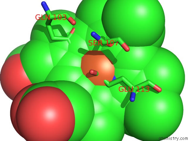
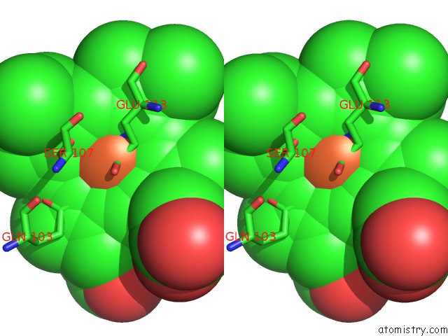
Iron binding site 1 out of 4 in 3sjl
Go back to
Iron binding site 1 out
of 4 in the Crystal Structure of the P107S-Maug/Pre-Methylamine Dehydrogenase Complex
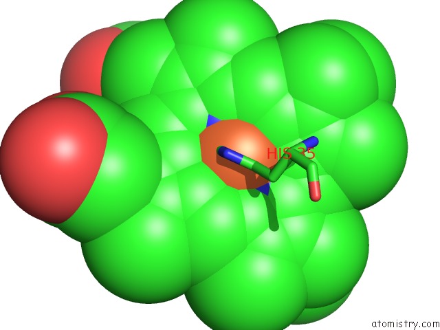
Mono view
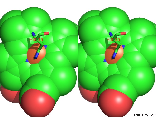
Stereo pair view
Mono view
Stereo pair view
A full contact list of Iron with other atoms in the Fe binding
site number 1 of Crystal Structure of the P107S-Maug/Pre-Methylamine Dehydrogenase Complex within 5.0Å range:
|
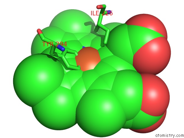
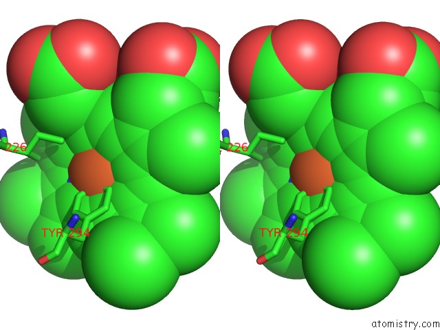
Iron binding site 2 out of 4 in 3sjl
Go back to
Iron binding site 2 out
of 4 in the Crystal Structure of the P107S-Maug/Pre-Methylamine Dehydrogenase Complex
Mono view
Stereo pair view
Mono view
Stereo pair view
A full contact list of Iron with other atoms in the Fe binding
site number 2 of Crystal Structure of the P107S-Maug/Pre-Methylamine Dehydrogenase Complex within 5.0Å range:
|
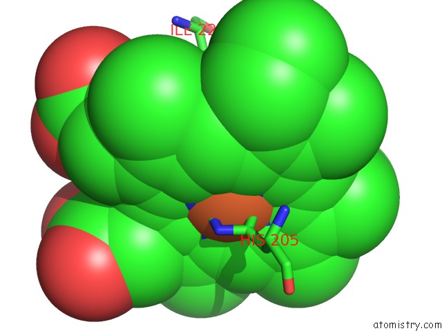
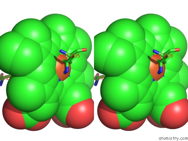
Iron binding site 3 out of 4 in 3sjl
Go back to
Iron binding site 3 out
of 4 in the Crystal Structure of the P107S-Maug/Pre-Methylamine Dehydrogenase Complex
Mono view
Stereo pair view
Mono view
Stereo pair view
A full contact list of Iron with other atoms in the Fe binding
site number 3 of Crystal Structure of the P107S-Maug/Pre-Methylamine Dehydrogenase Complex within 5.0Å range:
|
Iron binding site 4 out of 4 in 3sjl
Go back to
Iron binding site 4 out
of 4 in the Crystal Structure of the P107S-Maug/Pre-Methylamine Dehydrogenase Complex
Mono view
Stereo pair view
Mono view
Stereo pair view
A full contact list of Iron with other atoms in the Fe binding
site number 4 of Crystal Structure of the P107S-Maug/Pre-Methylamine Dehydrogenase Complex within 5.0Å range:
|
Reference:
M.Feng,
L.M.Jensen,
E.T.Yukl,
X.Wei,
A.Liu,
C.M.Wilmot,
V.L.Davidson.
Proline 107 Is A Major Determinant in Maintaining the Structure of the Distal Pocket and Reactivity of the High-Spin Heme of Maug. Biochemistry V. 51 1598 2012.
ISSN: ISSN 0006-2960
PubMed: 22299652
DOI: 10.1021/BI201882E
Page generated: Sun Aug 4 19:58:31 2024
ISSN: ISSN 0006-2960
PubMed: 22299652
DOI: 10.1021/BI201882E
Last articles
Zn in 9J0NZn in 9J0O
Zn in 9J0P
Zn in 9FJX
Zn in 9EKB
Zn in 9C0F
Zn in 9CAH
Zn in 9CH0
Zn in 9CH3
Zn in 9CH1