Iron »
PDB 3s6b-3sxt »
3swt »
Iron in PDB 3swt: Crystal Structure of the Taurine Catabolism Dioxygenase, Taud From Mycobacterium Marinum
Protein crystallography data
The structure of Crystal Structure of the Taurine Catabolism Dioxygenase, Taud From Mycobacterium Marinum, PDB code: 3swt
was solved by
Seattle Structural Genomics Center For Infectious Disease (Ssgcid),
with X-Ray Crystallography technique. A brief refinement statistics is given in the table below:
| Resolution Low / High (Å) | 50.00 / 2.05 |
| Space group | P 21 2 21 |
| Cell size a, b, c (Å), α, β, γ (°) | 69.098, 89.078, 104.703, 90.00, 90.00, 90.00 |
| R / Rfree (%) | 18.6 / 22.6 |
Iron Binding Sites:
The binding sites of Iron atom in the Crystal Structure of the Taurine Catabolism Dioxygenase, Taud From Mycobacterium Marinum
(pdb code 3swt). This binding sites where shown within
5.0 Angstroms radius around Iron atom.
In total 2 binding sites of Iron where determined in the Crystal Structure of the Taurine Catabolism Dioxygenase, Taud From Mycobacterium Marinum, PDB code: 3swt:
Jump to Iron binding site number: 1; 2;
In total 2 binding sites of Iron where determined in the Crystal Structure of the Taurine Catabolism Dioxygenase, Taud From Mycobacterium Marinum, PDB code: 3swt:
Jump to Iron binding site number: 1; 2;
Iron binding site 1 out of 2 in 3swt
Go back to
Iron binding site 1 out
of 2 in the Crystal Structure of the Taurine Catabolism Dioxygenase, Taud From Mycobacterium Marinum
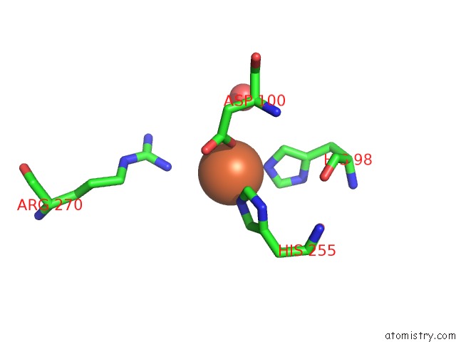
Mono view
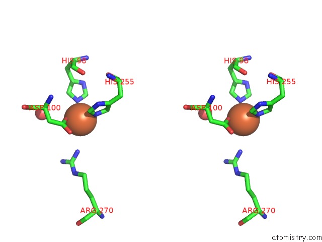
Stereo pair view
Mono view
Stereo pair view
A full contact list of Iron with other atoms in the Fe binding
site number 1 of Crystal Structure of the Taurine Catabolism Dioxygenase, Taud From Mycobacterium Marinum within 5.0Å range:
|
Iron binding site 2 out of 2 in 3swt
Go back to
Iron binding site 2 out
of 2 in the Crystal Structure of the Taurine Catabolism Dioxygenase, Taud From Mycobacterium Marinum
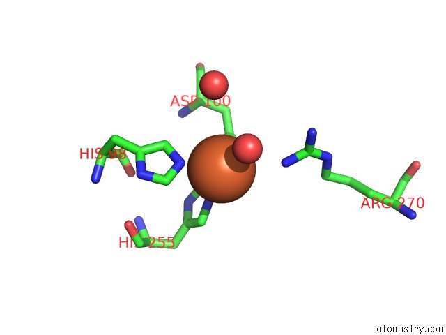
Mono view
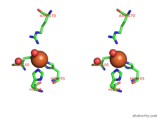
Stereo pair view
Mono view
Stereo pair view
A full contact list of Iron with other atoms in the Fe binding
site number 2 of Crystal Structure of the Taurine Catabolism Dioxygenase, Taud From Mycobacterium Marinum within 5.0Å range:
|
Reference:
L.Baugh,
I.Phan,
D.W.Begley,
M.C.Clifton,
B.Armour,
D.M.Dranow,
B.M.Taylor,
M.M.Muruthi,
J.Abendroth,
J.W.Fairman,
D.Fox,
S.H.Dieterich,
B.L.Staker,
A.S.Gardberg,
R.Choi,
S.N.Hewitt,
A.J.Napuli,
J.Myers,
L.K.Barrett,
Y.Zhang,
M.Ferrell,
E.Mundt,
K.Thompkins,
N.Tran,
S.Lyons-Abbott,
A.Abramov,
A.Sekar,
D.Serbzhinskiy,
D.Lorimer,
G.W.Buchko,
R.Stacy,
L.J.Stewart,
T.E.Edwards,
W.C.Van Voorhis,
P.J.Myler.
Increasing the Structural Coverage of Tuberculosis Drug Targets. Tuberculosis (Edinb) V. 95 142 2015.
ISSN: ISSN 1472-9792
PubMed: 25613812
DOI: 10.1016/J.TUBE.2014.12.003
Page generated: Sun Aug 4 20:04:29 2024
ISSN: ISSN 1472-9792
PubMed: 25613812
DOI: 10.1016/J.TUBE.2014.12.003
Last articles
Cl in 7Y6ICl in 7YBG
Cl in 7Y7Z
Cl in 7Y8I
Cl in 7Y7W
Cl in 7Y7Y
Cl in 7Y7V
Cl in 7Y5T
Cl in 7XXK
Cl in 7Y1W