Iron »
PDB 3sxv-3tgm »
3szk »
Iron in PDB 3szk: Crystal Structure of Human Methaemoglobin Complexed with the First Neat Domain of Isdh From Staphylococcus Aureus
Protein crystallography data
The structure of Crystal Structure of Human Methaemoglobin Complexed with the First Neat Domain of Isdh From Staphylococcus Aureus, PDB code: 3szk
was solved by
D.A.Jacques,
K.K.Kumar,
J.M.Guss,
D.A.Gell,
with X-Ray Crystallography technique. A brief refinement statistics is given in the table below:
| Resolution Low / High (Å) | 48.59 / 3.01 |
| Space group | P 21 21 21 |
| Cell size a, b, c (Å), α, β, γ (°) | 65.880, 123.206, 143.933, 90.00, 90.00, 90.00 |
| R / Rfree (%) | 24.5 / 27.5 |
Iron Binding Sites:
The binding sites of Iron atom in the Crystal Structure of Human Methaemoglobin Complexed with the First Neat Domain of Isdh From Staphylococcus Aureus
(pdb code 3szk). This binding sites where shown within
5.0 Angstroms radius around Iron atom.
In total 4 binding sites of Iron where determined in the Crystal Structure of Human Methaemoglobin Complexed with the First Neat Domain of Isdh From Staphylococcus Aureus, PDB code: 3szk:
Jump to Iron binding site number: 1; 2; 3; 4;
In total 4 binding sites of Iron where determined in the Crystal Structure of Human Methaemoglobin Complexed with the First Neat Domain of Isdh From Staphylococcus Aureus, PDB code: 3szk:
Jump to Iron binding site number: 1; 2; 3; 4;
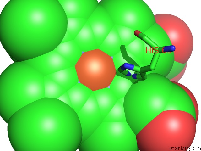
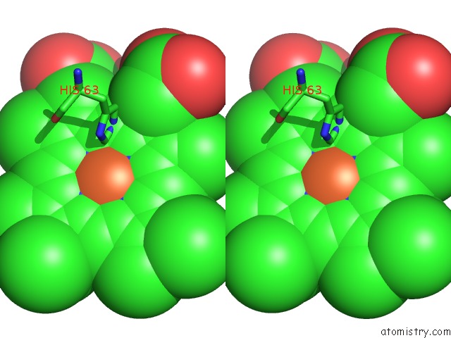
Iron binding site 1 out of 4 in 3szk
Go back to
Iron binding site 1 out
of 4 in the Crystal Structure of Human Methaemoglobin Complexed with the First Neat Domain of Isdh From Staphylococcus Aureus
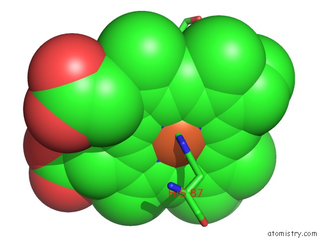
Mono view
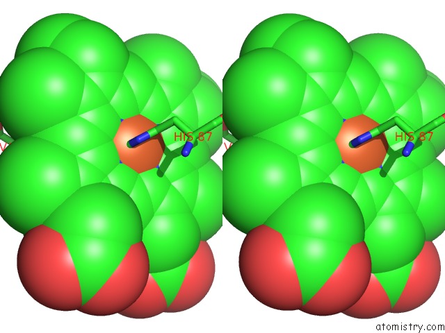
Stereo pair view
Mono view
Stereo pair view
A full contact list of Iron with other atoms in the Fe binding
site number 1 of Crystal Structure of Human Methaemoglobin Complexed with the First Neat Domain of Isdh From Staphylococcus Aureus within 5.0Å range:
|
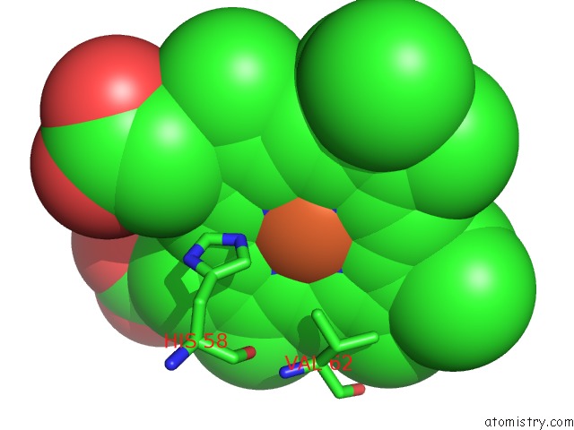
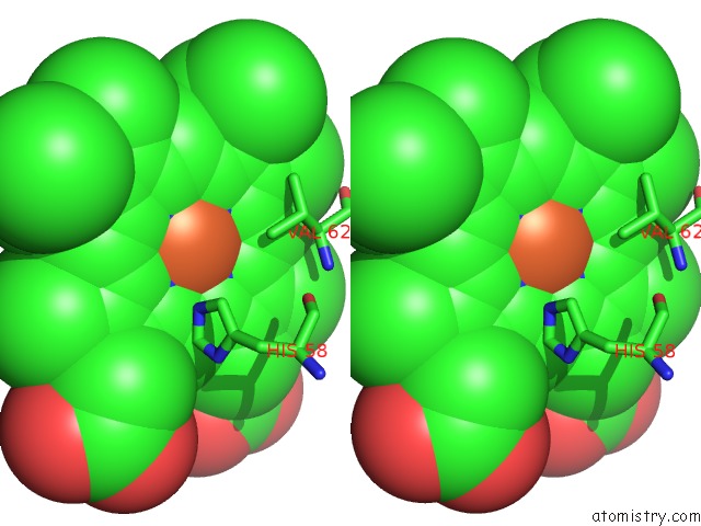
Iron binding site 2 out of 4 in 3szk
Go back to
Iron binding site 2 out
of 4 in the Crystal Structure of Human Methaemoglobin Complexed with the First Neat Domain of Isdh From Staphylococcus Aureus
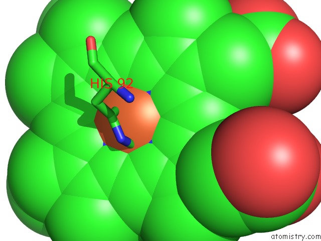
Mono view
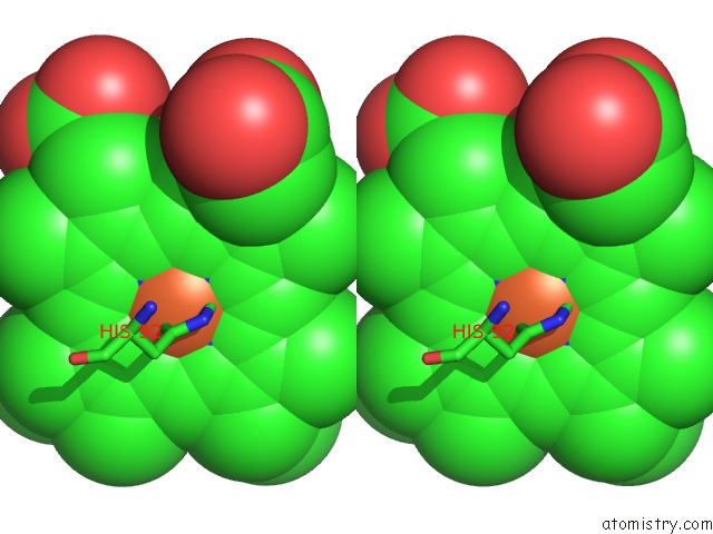
Stereo pair view
Mono view
Stereo pair view
A full contact list of Iron with other atoms in the Fe binding
site number 2 of Crystal Structure of Human Methaemoglobin Complexed with the First Neat Domain of Isdh From Staphylococcus Aureus within 5.0Å range:
|
Iron binding site 3 out of 4 in 3szk
Go back to
Iron binding site 3 out
of 4 in the Crystal Structure of Human Methaemoglobin Complexed with the First Neat Domain of Isdh From Staphylococcus Aureus
Mono view
Stereo pair view
Mono view
Stereo pair view
A full contact list of Iron with other atoms in the Fe binding
site number 3 of Crystal Structure of Human Methaemoglobin Complexed with the First Neat Domain of Isdh From Staphylococcus Aureus within 5.0Å range:
|
Iron binding site 4 out of 4 in 3szk
Go back to
Iron binding site 4 out
of 4 in the Crystal Structure of Human Methaemoglobin Complexed with the First Neat Domain of Isdh From Staphylococcus Aureus
Mono view
Stereo pair view
Mono view
Stereo pair view
A full contact list of Iron with other atoms in the Fe binding
site number 4 of Crystal Structure of Human Methaemoglobin Complexed with the First Neat Domain of Isdh From Staphylococcus Aureus within 5.0Å range:
|
Reference:
K.Krishna Kumar,
D.A.Jacques,
G.Pishchany,
T.Caradoc-Davies,
T.Spirig,
G.R.Malmirchegini,
D.B.Langley,
C.F.Dickson,
J.P.Mackay,
R.T.Clubb,
E.P.Skaar,
J.M.Guss,
D.A.Gell.
Structural Basis For Hemoglobin Capture By Staphylococcus Aureus Cell-Surface Protein, Isdh J.Biol.Chem. V. 286 38439 2011.
ISSN: ISSN 0021-9258
PubMed: 21917915
DOI: 10.1074/JBC.M111.287300
Page generated: Sun Aug 4 20:12:30 2024
ISSN: ISSN 0021-9258
PubMed: 21917915
DOI: 10.1074/JBC.M111.287300
Last articles
Zn in 9MJ5Zn in 9HNW
Zn in 9G0L
Zn in 9FNE
Zn in 9DZN
Zn in 9E0I
Zn in 9D32
Zn in 9DAK
Zn in 8ZXC
Zn in 8ZUF