Iron »
PDB 3sxv-3tgm »
3szu »
Iron in PDB 3szu: Isph:Hmbpp Complex Structure of E126Q Mutant
Enzymatic activity of Isph:Hmbpp Complex Structure of E126Q Mutant
All present enzymatic activity of Isph:Hmbpp Complex Structure of E126Q Mutant:
1.17.1.2;
1.17.1.2;
Protein crystallography data
The structure of Isph:Hmbpp Complex Structure of E126Q Mutant, PDB code: 3szu
was solved by
I.Span,
T.Graewert,
A.Bacher,
W.Eisenreich,
M.Groll,
with X-Ray Crystallography technique. A brief refinement statistics is given in the table below:
| Resolution Low / High (Å) | 10.00 / 1.40 |
| Space group | P 21 21 21 |
| Cell size a, b, c (Å), α, β, γ (°) | 70.190, 80.390, 112.010, 90.00, 90.00, 90.00 |
| R / Rfree (%) | 17.5 / 21.5 |
Iron Binding Sites:
The binding sites of Iron atom in the Isph:Hmbpp Complex Structure of E126Q Mutant
(pdb code 3szu). This binding sites where shown within
5.0 Angstroms radius around Iron atom.
In total 6 binding sites of Iron where determined in the Isph:Hmbpp Complex Structure of E126Q Mutant, PDB code: 3szu:
Jump to Iron binding site number: 1; 2; 3; 4; 5; 6;
In total 6 binding sites of Iron where determined in the Isph:Hmbpp Complex Structure of E126Q Mutant, PDB code: 3szu:
Jump to Iron binding site number: 1; 2; 3; 4; 5; 6;
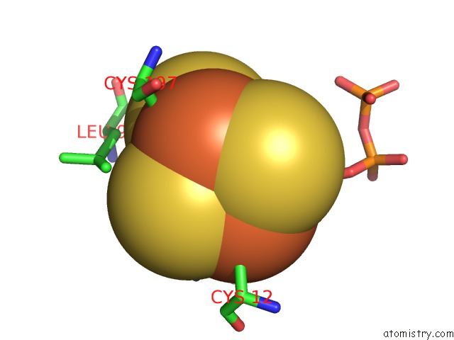
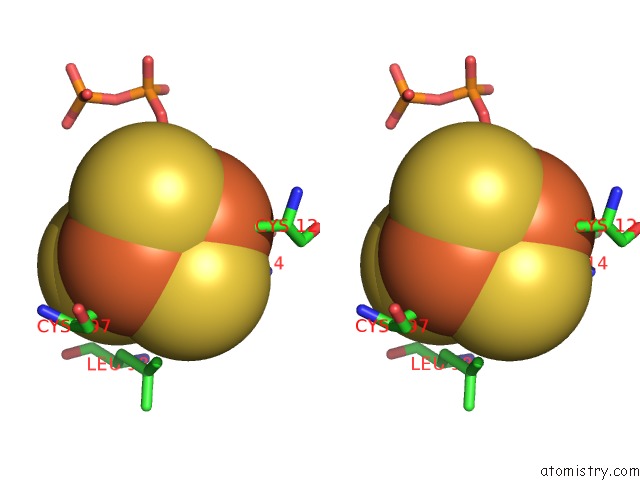
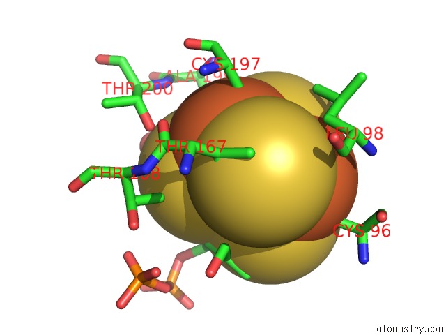
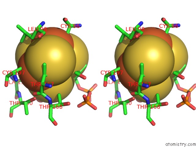
Iron binding site 1 out of 6 in 3szu
Go back to
Iron binding site 1 out
of 6 in the Isph:Hmbpp Complex Structure of E126Q Mutant
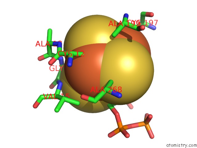
Mono view
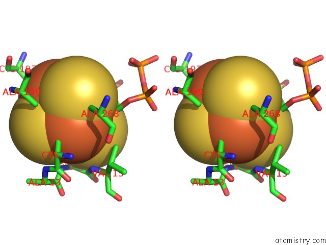
Stereo pair view
Mono view
Stereo pair view
A full contact list of Iron with other atoms in the Fe binding
site number 1 of Isph:Hmbpp Complex Structure of E126Q Mutant within 5.0Å range:
|
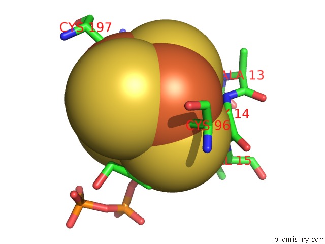
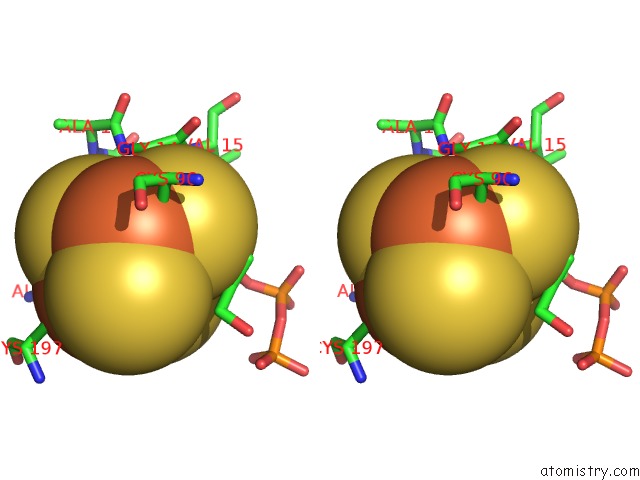
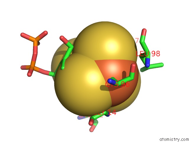
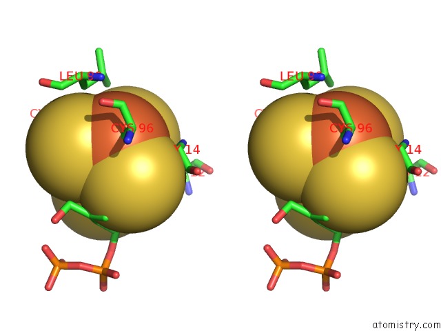
Iron binding site 2 out of 6 in 3szu
Go back to
Iron binding site 2 out
of 6 in the Isph:Hmbpp Complex Structure of E126Q Mutant
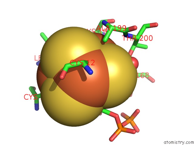
Mono view
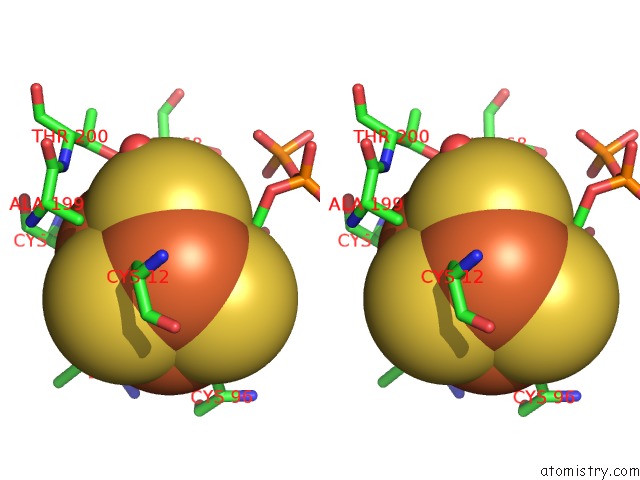
Stereo pair view
Mono view
Stereo pair view
A full contact list of Iron with other atoms in the Fe binding
site number 2 of Isph:Hmbpp Complex Structure of E126Q Mutant within 5.0Å range:
|
Iron binding site 3 out of 6 in 3szu
Go back to
Iron binding site 3 out
of 6 in the Isph:Hmbpp Complex Structure of E126Q Mutant
Mono view
Stereo pair view
Mono view
Stereo pair view
A full contact list of Iron with other atoms in the Fe binding
site number 3 of Isph:Hmbpp Complex Structure of E126Q Mutant within 5.0Å range:
|
Iron binding site 4 out of 6 in 3szu
Go back to
Iron binding site 4 out
of 6 in the Isph:Hmbpp Complex Structure of E126Q Mutant
Mono view
Stereo pair view
Mono view
Stereo pair view
A full contact list of Iron with other atoms in the Fe binding
site number 4 of Isph:Hmbpp Complex Structure of E126Q Mutant within 5.0Å range:
|
Iron binding site 5 out of 6 in 3szu
Go back to
Iron binding site 5 out
of 6 in the Isph:Hmbpp Complex Structure of E126Q Mutant
Mono view
Stereo pair view
Mono view
Stereo pair view
A full contact list of Iron with other atoms in the Fe binding
site number 5 of Isph:Hmbpp Complex Structure of E126Q Mutant within 5.0Å range:
|
Iron binding site 6 out of 6 in 3szu
Go back to
Iron binding site 6 out
of 6 in the Isph:Hmbpp Complex Structure of E126Q Mutant
Mono view
Stereo pair view
Mono view
Stereo pair view
A full contact list of Iron with other atoms in the Fe binding
site number 6 of Isph:Hmbpp Complex Structure of E126Q Mutant within 5.0Å range:
|
Reference:
I.Span,
T.Grawert,
A.Bacher,
W.Eisenreich,
M.Groll.
Crystal Structures of Mutant Isph Proteins Reveal A Rotation of the Substrate'S Hydroxymethyl Group During Catalysis. J.Mol.Biol. V. 416 1 2012.
ISSN: ISSN 0022-2836
PubMed: 22137895
DOI: 10.1016/J.JMB.2011.11.033
Page generated: Sun Aug 4 20:12:30 2024
ISSN: ISSN 0022-2836
PubMed: 22137895
DOI: 10.1016/J.JMB.2011.11.033
Last articles
Zn in 9MJ5Zn in 9HNW
Zn in 9G0L
Zn in 9FNE
Zn in 9DZN
Zn in 9E0I
Zn in 9D32
Zn in 9DAK
Zn in 8ZXC
Zn in 8ZUF