Iron »
PDB 3sxv-3tgm »
3t4v »
Iron in PDB 3t4v: Crystal Structure of Alkb in Complex with Fe(III) and N-Oxalyl-S-(2- Napthalenemethyl)-L-Cysteine
Protein crystallography data
The structure of Crystal Structure of Alkb in Complex with Fe(III) and N-Oxalyl-S-(2- Napthalenemethyl)-L-Cysteine, PDB code: 3t4v
was solved by
W.S.Aik,
M.A.Mcdonough,
C.J.Schofield,
with X-Ray Crystallography technique. A brief refinement statistics is given in the table below:
| Resolution Low / High (Å) | 22.99 / 1.73 |
| Space group | P 1 |
| Cell size a, b, c (Å), α, β, γ (°) | 37.070, 39.234, 40.261, 76.33, 74.56, 65.28 |
| R / Rfree (%) | 16.1 / 20.2 |
Iron Binding Sites:
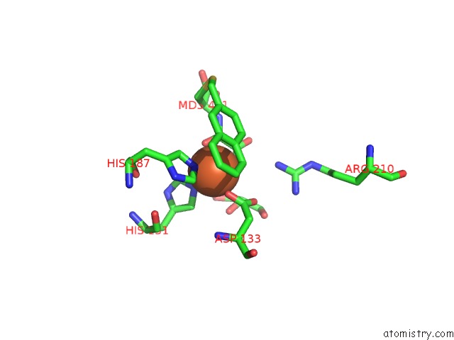
The binding sites of Iron atom in the Crystal Structure of Alkb in Complex with Fe(III) and N-Oxalyl-S-(2- Napthalenemethyl)-L-Cysteine
(pdb code 3t4v). This binding sites where shown within
5.0 Angstroms radius around Iron atom.
In total only one binding site of Iron was determined in the Crystal Structure of Alkb in Complex with Fe(III) and N-Oxalyl-S-(2- Napthalenemethyl)-L-Cysteine, PDB code: 3t4v:
In total only one binding site of Iron was determined in the Crystal Structure of Alkb in Complex with Fe(III) and N-Oxalyl-S-(2- Napthalenemethyl)-L-Cysteine, PDB code: 3t4v:
Iron binding site 1 out of 1 in 3t4v
Go back to
Iron binding site 1 out
of 1 in the Crystal Structure of Alkb in Complex with Fe(III) and N-Oxalyl-S-(2- Napthalenemethyl)-L-Cysteine
Mono view
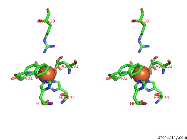
Stereo pair view
Mono view
Stereo pair view
A full contact list of Iron with other atoms in the Fe binding
site number 1 of Crystal Structure of Alkb in Complex with Fe(III) and N-Oxalyl-S-(2- Napthalenemethyl)-L-Cysteine within 5.0Å range:
|
Reference:
E.C.Woon,
M.Demetriades,
E.A.Bagg,
W.Aik,
S.M.Krylova,
J.H.Ma,
M.Chan,
L.J.Walport,
D.W.Wegman,
K.N.Dack,
M.A.Mcdonough,
S.N.Krylov,
C.J.Schofield.
Dynamic Combinatorial Mass Spectrometry Leads to Inhibitors of A 2-Oxoglutarate-Dependent Nucleic Acid Demethylase. J.Med.Chem. V. 55 2173 2012.
ISSN: ISSN 0022-2623
PubMed: 22263962
DOI: 10.1021/JM201417E
Page generated: Sun Aug 4 20:17:04 2024
ISSN: ISSN 0022-2623
PubMed: 22263962
DOI: 10.1021/JM201417E
Last articles
Zn in 9J0NZn in 9J0O
Zn in 9J0P
Zn in 9FJX
Zn in 9EKB
Zn in 9C0F
Zn in 9CAH
Zn in 9CH0
Zn in 9CH3
Zn in 9CH1