Iron »
PDB 3sxv-3tgm »
3t81 »
Iron in PDB 3t81: Crystal Structure of Diiron Adenine Deaminase
Enzymatic activity of Crystal Structure of Diiron Adenine Deaminase
All present enzymatic activity of Crystal Structure of Diiron Adenine Deaminase:
3.5.4.2;
3.5.4.2;
Protein crystallography data
The structure of Crystal Structure of Diiron Adenine Deaminase, PDB code: 3t81
was solved by
A.Bagaria,
D.Kumaran,
S.K.Burley,
S.Swaminathan,
New York Sgx Researchcenter For Structural Genomics (Nysgxrc),
with X-Ray Crystallography technique. A brief refinement statistics is given in the table below:
| Resolution Low / High (Å) | 48.89 / 2.63 |
| Space group | P 1 21 1 |
| Cell size a, b, c (Å), α, β, γ (°) | 61.662, 131.845, 69.628, 90.00, 97.04, 90.00 |
| R / Rfree (%) | 18.5 / 28.7 |
Iron Binding Sites:
The binding sites of Iron atom in the Crystal Structure of Diiron Adenine Deaminase
(pdb code 3t81). This binding sites where shown within
5.0 Angstroms radius around Iron atom.
In total 6 binding sites of Iron where determined in the Crystal Structure of Diiron Adenine Deaminase, PDB code: 3t81:
Jump to Iron binding site number: 1; 2; 3; 4; 5; 6;
In total 6 binding sites of Iron where determined in the Crystal Structure of Diiron Adenine Deaminase, PDB code: 3t81:
Jump to Iron binding site number: 1; 2; 3; 4; 5; 6;
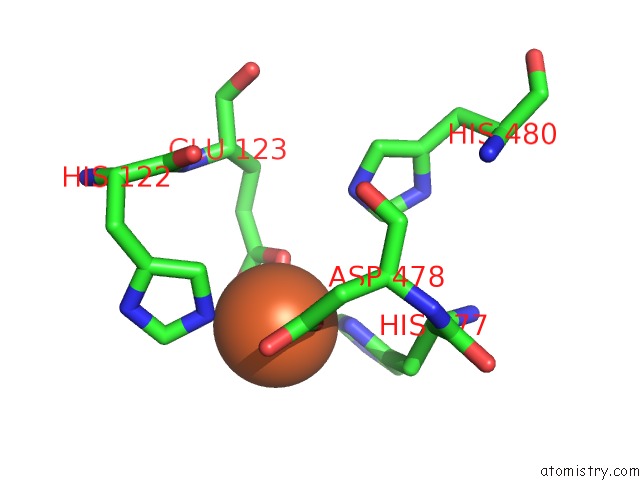
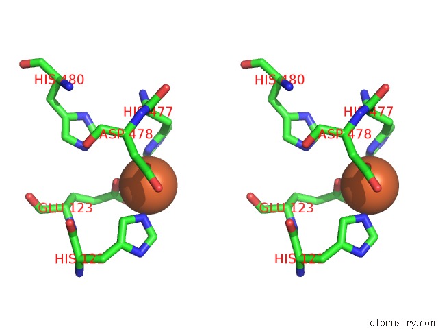
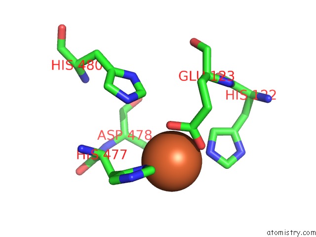
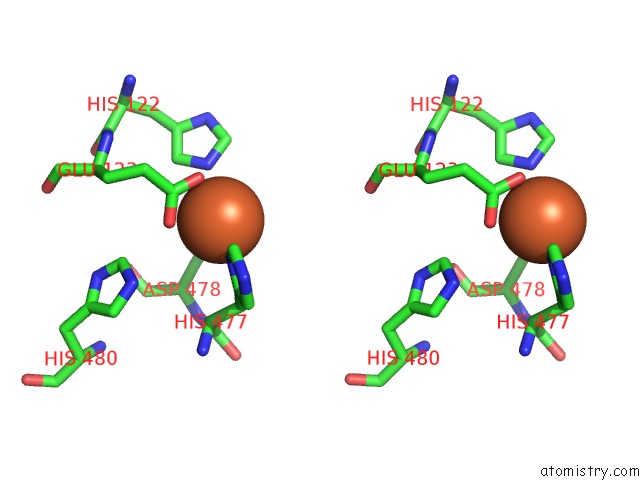
Iron binding site 1 out of 6 in 3t81
Go back to
Iron binding site 1 out
of 6 in the Crystal Structure of Diiron Adenine Deaminase
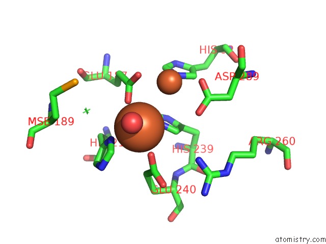
Mono view
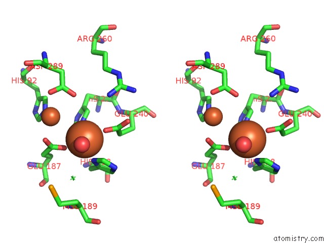
Stereo pair view
Mono view
Stereo pair view
A full contact list of Iron with other atoms in the Fe binding
site number 1 of Crystal Structure of Diiron Adenine Deaminase within 5.0Å range:
|
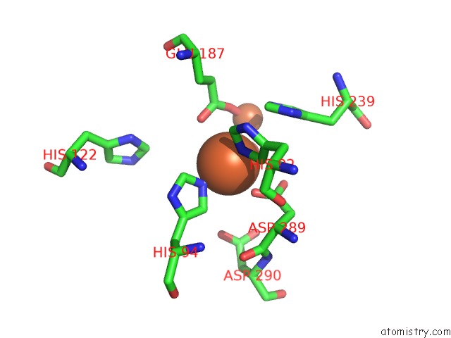
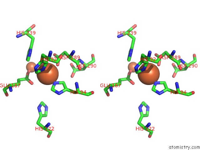
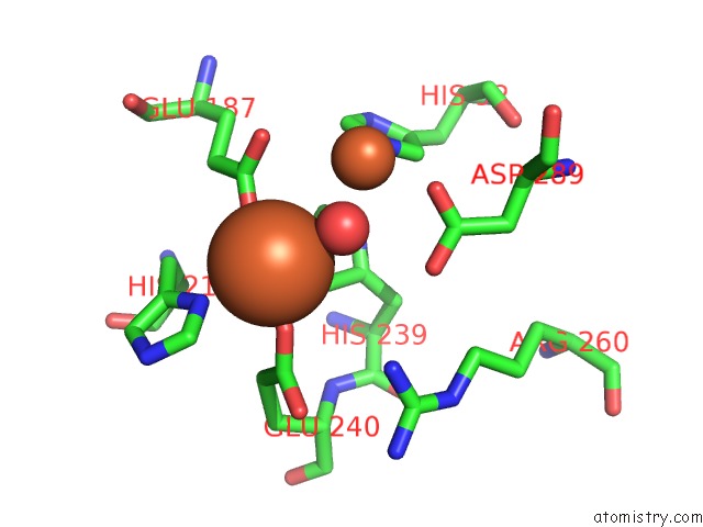
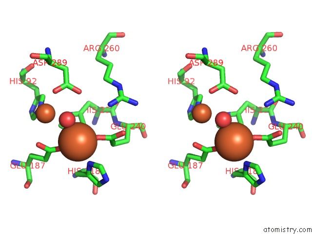
Iron binding site 2 out of 6 in 3t81
Go back to
Iron binding site 2 out
of 6 in the Crystal Structure of Diiron Adenine Deaminase
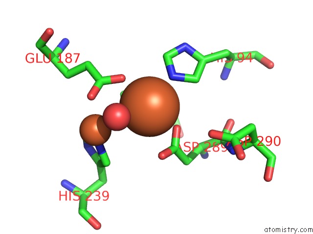
Mono view
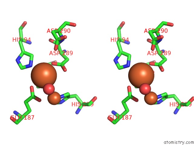
Stereo pair view
Mono view
Stereo pair view
A full contact list of Iron with other atoms in the Fe binding
site number 2 of Crystal Structure of Diiron Adenine Deaminase within 5.0Å range:
|
Iron binding site 3 out of 6 in 3t81
Go back to
Iron binding site 3 out
of 6 in the Crystal Structure of Diiron Adenine Deaminase
Mono view
Stereo pair view
Mono view
Stereo pair view
A full contact list of Iron with other atoms in the Fe binding
site number 3 of Crystal Structure of Diiron Adenine Deaminase within 5.0Å range:
|
Iron binding site 4 out of 6 in 3t81
Go back to
Iron binding site 4 out
of 6 in the Crystal Structure of Diiron Adenine Deaminase
Mono view
Stereo pair view
Mono view
Stereo pair view
A full contact list of Iron with other atoms in the Fe binding
site number 4 of Crystal Structure of Diiron Adenine Deaminase within 5.0Å range:
|
Iron binding site 5 out of 6 in 3t81
Go back to
Iron binding site 5 out
of 6 in the Crystal Structure of Diiron Adenine Deaminase
Mono view
Stereo pair view
Mono view
Stereo pair view
A full contact list of Iron with other atoms in the Fe binding
site number 5 of Crystal Structure of Diiron Adenine Deaminase within 5.0Å range:
|
Iron binding site 6 out of 6 in 3t81
Go back to
Iron binding site 6 out
of 6 in the Crystal Structure of Diiron Adenine Deaminase
Mono view
Stereo pair view
Mono view
Stereo pair view
A full contact list of Iron with other atoms in the Fe binding
site number 6 of Crystal Structure of Diiron Adenine Deaminase within 5.0Å range:
|
Reference:
A.Bagaria,
D.Kumaran,
S.K.Burley,
S.Swaminathan.
Crystal Structure of Diiron Adenine Deaminase To Be Published.
Page generated: Sun Aug 4 20:19:44 2024
Last articles
F in 7R9CF in 7QZ7
F in 7R9F
F in 7R7R
F in 7R53
F in 7R3V
F in 7R7K
F in 7QZ9
F in 7QZ8
F in 7R2B