Iron »
PDB 3tgu-3u3e »
3tld »
Iron in PDB 3tld: Crystal Structure of Y29F Mutant of Vitreoscilla Hemoglobin
Protein crystallography data
The structure of Crystal Structure of Y29F Mutant of Vitreoscilla Hemoglobin, PDB code: 3tld
was solved by
S.Ratakonda,
A.Anand,
K.Dikshit,
B.C.Stark,
A.J.Howard,
with X-Ray Crystallography technique. A brief refinement statistics is given in the table below:
| Resolution Low / High (Å) | 31.57 / 1.90 |
| Space group | P 1 21 1 |
| Cell size a, b, c (Å), α, β, γ (°) | 61.614, 41.068, 62.395, 90.00, 105.58, 90.00 |
| R / Rfree (%) | 16.5 / 18.9 |
Iron Binding Sites:
The binding sites of Iron atom in the Crystal Structure of Y29F Mutant of Vitreoscilla Hemoglobin
(pdb code 3tld). This binding sites where shown within
5.0 Angstroms radius around Iron atom.
In total 2 binding sites of Iron where determined in the Crystal Structure of Y29F Mutant of Vitreoscilla Hemoglobin, PDB code: 3tld:
Jump to Iron binding site number: 1; 2;
In total 2 binding sites of Iron where determined in the Crystal Structure of Y29F Mutant of Vitreoscilla Hemoglobin, PDB code: 3tld:
Jump to Iron binding site number: 1; 2;
Iron binding site 1 out of 2 in 3tld
Go back to
Iron binding site 1 out
of 2 in the Crystal Structure of Y29F Mutant of Vitreoscilla Hemoglobin
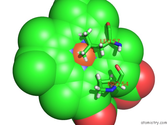
Mono view
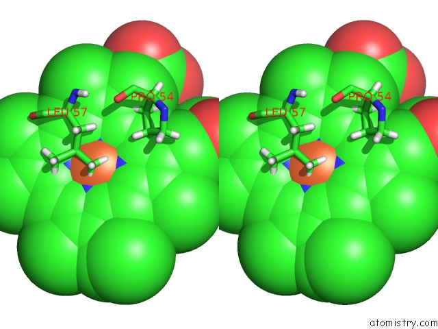
Stereo pair view
Mono view
Stereo pair view
A full contact list of Iron with other atoms in the Fe binding
site number 1 of Crystal Structure of Y29F Mutant of Vitreoscilla Hemoglobin within 5.0Å range:
|
Iron binding site 2 out of 2 in 3tld
Go back to
Iron binding site 2 out
of 2 in the Crystal Structure of Y29F Mutant of Vitreoscilla Hemoglobin
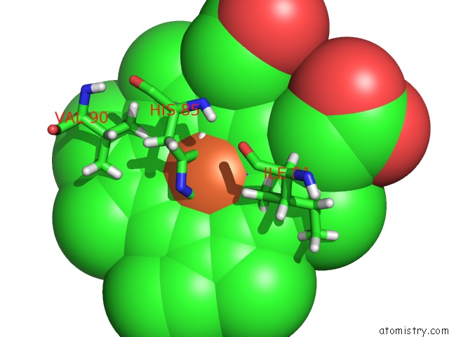
Mono view
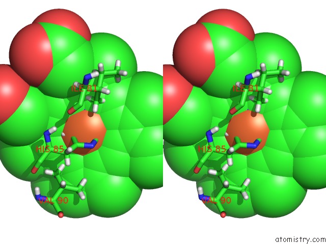
Stereo pair view
Mono view
Stereo pair view
A full contact list of Iron with other atoms in the Fe binding
site number 2 of Crystal Structure of Y29F Mutant of Vitreoscilla Hemoglobin within 5.0Å range:
|
Reference:
S.Ratakonda,
A.Anand,
K.Dikshit,
B.C.Stark,
A.J.Howard.
Crystallographic Structure Determination of B10 Mutants of Vitreoscilla Hemoglobin: Role of TYR29 (B10) in the Structure of the Ligand-Binding Site. Acta Crystallogr.,Sect.F V. 69 215 2013.
ISSN: ESSN 1744-3091
PubMed: 23519792
DOI: 10.1107/S1744309112044818
Page generated: Sun Aug 4 20:28:30 2024
ISSN: ESSN 1744-3091
PubMed: 23519792
DOI: 10.1107/S1744309112044818
Last articles
Cl in 5WQ3Cl in 5WRO
Cl in 5WRB
Cl in 5WRA
Cl in 5WR9
Cl in 5WO3
Cl in 5WO4
Cl in 5WPN
Cl in 5WO2
Cl in 5WO1