Iron »
PDB 3tgu-3u3e »
3tmz »
Iron in PDB 3tmz: Crystal Structure of P450 2B4(H226Y) in Complex with Amlodipine
Enzymatic activity of Crystal Structure of P450 2B4(H226Y) in Complex with Amlodipine
All present enzymatic activity of Crystal Structure of P450 2B4(H226Y) in Complex with Amlodipine:
1.14.14.1;
1.14.14.1;
Protein crystallography data
The structure of Crystal Structure of P450 2B4(H226Y) in Complex with Amlodipine, PDB code: 3tmz
was solved by
M.B.Shah,
J.Pascual,
C.D.Stout,
J.R.Halpert,
with X-Ray Crystallography technique. A brief refinement statistics is given in the table below:
| Resolution Low / High (Å) | 38.93 / 2.25 |
| Space group | P 31 2 1 |
| Cell size a, b, c (Å), α, β, γ (°) | 92.995, 92.995, 152.773, 90.00, 90.00, 120.00 |
| R / Rfree (%) | 19.1 / 25.1 |
Other elements in 3tmz:
The structure of Crystal Structure of P450 2B4(H226Y) in Complex with Amlodipine also contains other interesting chemical elements:
| Chlorine | (Cl) | 3 atoms |
Iron Binding Sites:
The binding sites of Iron atom in the Crystal Structure of P450 2B4(H226Y) in Complex with Amlodipine
(pdb code 3tmz). This binding sites where shown within
5.0 Angstroms radius around Iron atom.
In total only one binding site of Iron was determined in the Crystal Structure of P450 2B4(H226Y) in Complex with Amlodipine, PDB code: 3tmz:
In total only one binding site of Iron was determined in the Crystal Structure of P450 2B4(H226Y) in Complex with Amlodipine, PDB code: 3tmz:
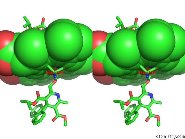
Iron binding site 1 out of 1 in 3tmz
Go back to
Iron binding site 1 out
of 1 in the Crystal Structure of P450 2B4(H226Y) in Complex with Amlodipine
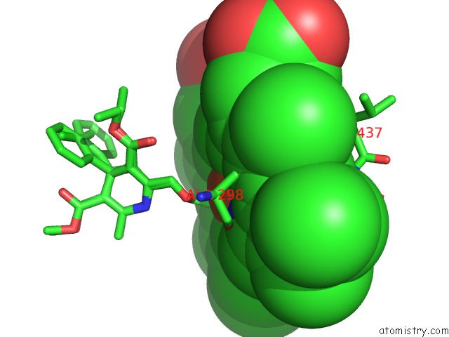
Mono view
Stereo pair view
Mono view
Stereo pair view
A full contact list of Iron with other atoms in the Fe binding
site number 1 of Crystal Structure of P450 2B4(H226Y) in Complex with Amlodipine within 5.0Å range:
|
Reference:
M.B.Shah,
P.R.Wilderman,
J.Pascual,
Q.Zhang,
C.D.Stout,
J.R.Halpert.
Conformational Adaptation of Human Cytochrome P450 2B6 and Rabbit Cytochrome P450 2B4 Revealed Upon Binding Multiple Amlodipine Molecules. Biochemistry V. 51 7225 2012.
ISSN: ISSN 0006-2960
PubMed: 22909231
DOI: 10.1021/BI300894Z
Page generated: Sun Aug 4 20:31:40 2024
ISSN: ISSN 0006-2960
PubMed: 22909231
DOI: 10.1021/BI300894Z
Last articles
Zn in 9J0NZn in 9J0O
Zn in 9J0P
Zn in 9FJX
Zn in 9EKB
Zn in 9C0F
Zn in 9CAH
Zn in 9CH0
Zn in 9CH3
Zn in 9CH1