Iron »
PDB 3tgu-3u3e »
3tqj »
Iron in PDB 3tqj: Structure of the Superoxide Dismutase (Fe) (Sodb) From Coxiella Burnetii
Enzymatic activity of Structure of the Superoxide Dismutase (Fe) (Sodb) From Coxiella Burnetii
All present enzymatic activity of Structure of the Superoxide Dismutase (Fe) (Sodb) From Coxiella Burnetii:
1.15.1.1;
1.15.1.1;
Protein crystallography data
The structure of Structure of the Superoxide Dismutase (Fe) (Sodb) From Coxiella Burnetii, PDB code: 3tqj
was solved by
M.C.Franklin,
J.Cheung,
M.Cassidy,
J.Love,
with X-Ray Crystallography technique. A brief refinement statistics is given in the table below:
| Resolution Low / High (Å) | 56.15 / 2.00 |
| Space group | P 21 21 21 |
| Cell size a, b, c (Å), α, β, γ (°) | 60.102, 65.285, 110.069, 90.00, 90.00, 90.00 |
| R / Rfree (%) | 23.3 / 27.1 |
Iron Binding Sites:
The binding sites of Iron atom in the Structure of the Superoxide Dismutase (Fe) (Sodb) From Coxiella Burnetii
(pdb code 3tqj). This binding sites where shown within
5.0 Angstroms radius around Iron atom.
In total 2 binding sites of Iron where determined in the Structure of the Superoxide Dismutase (Fe) (Sodb) From Coxiella Burnetii, PDB code: 3tqj:
Jump to Iron binding site number: 1; 2;
In total 2 binding sites of Iron where determined in the Structure of the Superoxide Dismutase (Fe) (Sodb) From Coxiella Burnetii, PDB code: 3tqj:
Jump to Iron binding site number: 1; 2;
Iron binding site 1 out of 2 in 3tqj
Go back to
Iron binding site 1 out
of 2 in the Structure of the Superoxide Dismutase (Fe) (Sodb) From Coxiella Burnetii
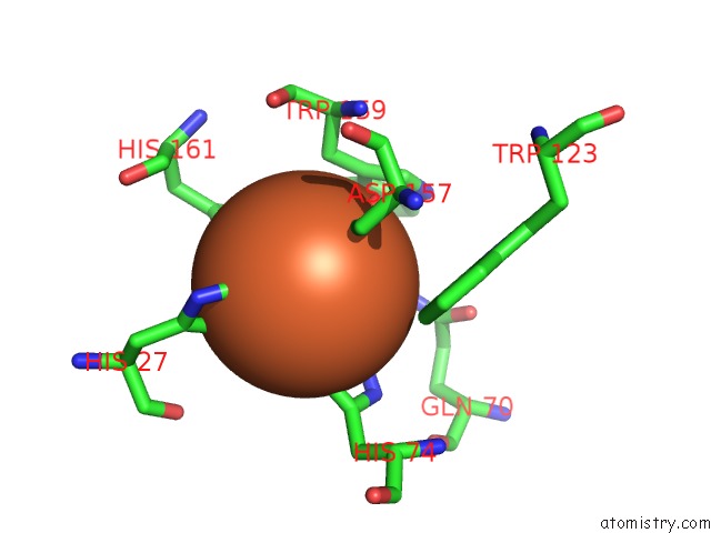
Mono view
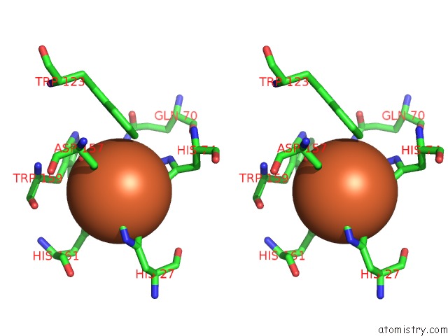
Stereo pair view
Mono view
Stereo pair view
A full contact list of Iron with other atoms in the Fe binding
site number 1 of Structure of the Superoxide Dismutase (Fe) (Sodb) From Coxiella Burnetii within 5.0Å range:
|
Iron binding site 2 out of 2 in 3tqj
Go back to
Iron binding site 2 out
of 2 in the Structure of the Superoxide Dismutase (Fe) (Sodb) From Coxiella Burnetii
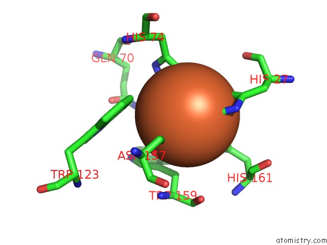
Mono view
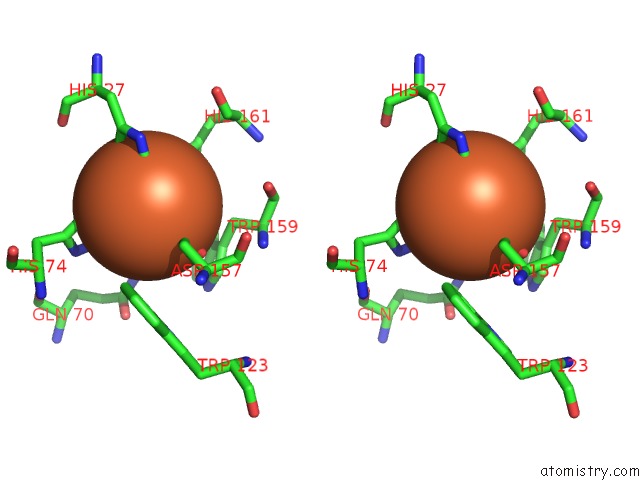
Stereo pair view
Mono view
Stereo pair view
A full contact list of Iron with other atoms in the Fe binding
site number 2 of Structure of the Superoxide Dismutase (Fe) (Sodb) From Coxiella Burnetii within 5.0Å range:
|
Reference:
M.C.Franklin,
J.Cheung,
M.J.Rudolph,
F.Burshteyn,
M.Cassidy,
E.Gary,
B.Hillerich,
Z.K.Yao,
P.R.Carlier,
M.Totrov,
J.D.Love.
Structural Genomics For Drug Design Against the Pathogen Coxiella Burnetii. Proteins V. 83 2124 2015.
ISSN: ISSN 0887-3585
PubMed: 26033498
DOI: 10.1002/PROT.24841
Page generated: Sun Aug 4 20:33:11 2024
ISSN: ISSN 0887-3585
PubMed: 26033498
DOI: 10.1002/PROT.24841
Last articles
Fe in 2YXOFe in 2YRS
Fe in 2YXC
Fe in 2YNM
Fe in 2YVJ
Fe in 2YP1
Fe in 2YU2
Fe in 2YU1
Fe in 2YQB
Fe in 2YOO