Iron »
PDB 3uhg-3v2z »
3unc »
Iron in PDB 3unc: Crystal Structure of Bovine Milk Xanthine Dehydrogenase to 1.65A Resolution
Enzymatic activity of Crystal Structure of Bovine Milk Xanthine Dehydrogenase to 1.65A Resolution
All present enzymatic activity of Crystal Structure of Bovine Milk Xanthine Dehydrogenase to 1.65A Resolution:
1.17.1.4; 1.17.3.2;
1.17.1.4; 1.17.3.2;
Protein crystallography data
The structure of Crystal Structure of Bovine Milk Xanthine Dehydrogenase to 1.65A Resolution, PDB code: 3unc
was solved by
B.T.Eger,
K.Okamoto,
T.Nishino,
E.F.Pai,
with X-Ray Crystallography technique. A brief refinement statistics is given in the table below:
| Resolution Low / High (Å) | 20.00 / 1.65 |
| Space group | C 1 2 1 |
| Cell size a, b, c (Å), α, β, γ (°) | 167.573, 124.370, 148.177, 90.00, 91.02, 90.00 |
| R / Rfree (%) | 17.7 / 19.6 |
Other elements in 3unc:
The structure of Crystal Structure of Bovine Milk Xanthine Dehydrogenase to 1.65A Resolution also contains other interesting chemical elements:
| Molybdenum | (Mo) | 2 atoms |
| Calcium | (Ca) | 2 atoms |
Iron Binding Sites:
The binding sites of Iron atom in the Crystal Structure of Bovine Milk Xanthine Dehydrogenase to 1.65A Resolution
(pdb code 3unc). This binding sites where shown within
5.0 Angstroms radius around Iron atom.
In total 8 binding sites of Iron where determined in the Crystal Structure of Bovine Milk Xanthine Dehydrogenase to 1.65A Resolution, PDB code: 3unc:
Jump to Iron binding site number: 1; 2; 3; 4; 5; 6; 7; 8;
In total 8 binding sites of Iron where determined in the Crystal Structure of Bovine Milk Xanthine Dehydrogenase to 1.65A Resolution, PDB code: 3unc:
Jump to Iron binding site number: 1; 2; 3; 4; 5; 6; 7; 8;
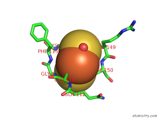
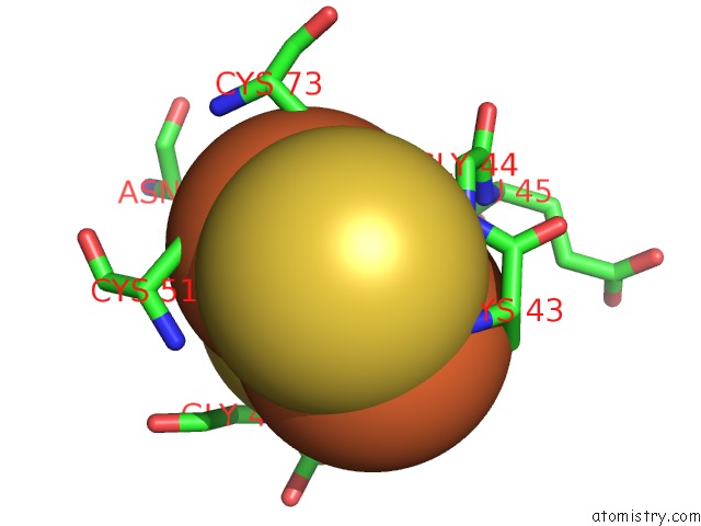
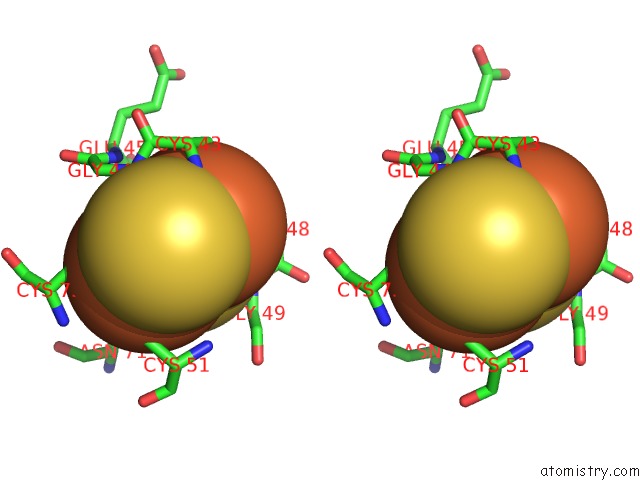
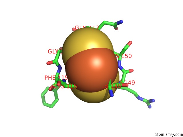
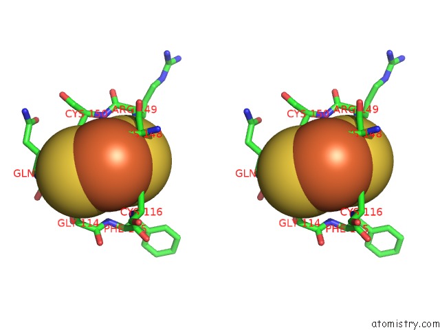
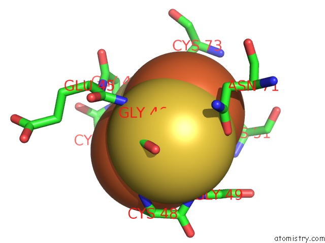
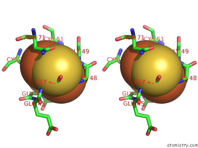
Iron binding site 1 out of 8 in 3unc
Go back to
Iron binding site 1 out
of 8 in the Crystal Structure of Bovine Milk Xanthine Dehydrogenase to 1.65A Resolution
Mono view
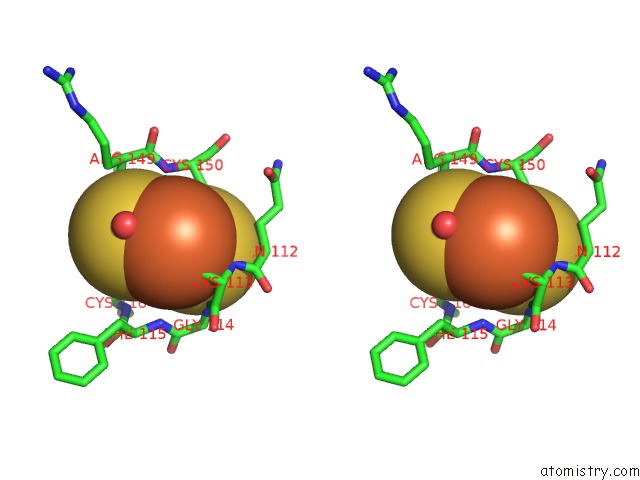
Stereo pair view
Mono view
Stereo pair view
A full contact list of Iron with other atoms in the Fe binding
site number 1 of Crystal Structure of Bovine Milk Xanthine Dehydrogenase to 1.65A Resolution within 5.0Å range:
|
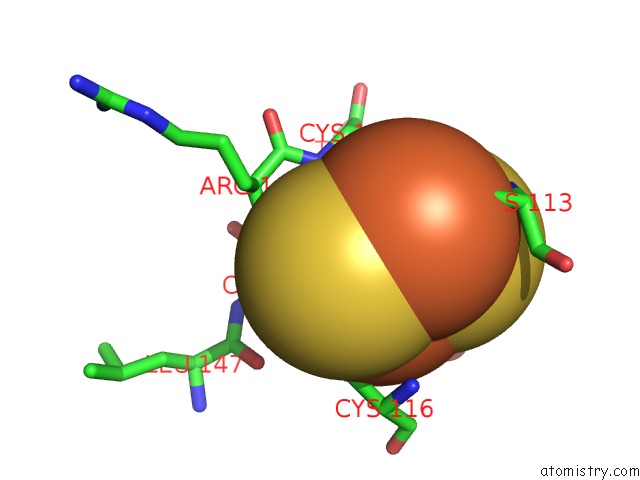
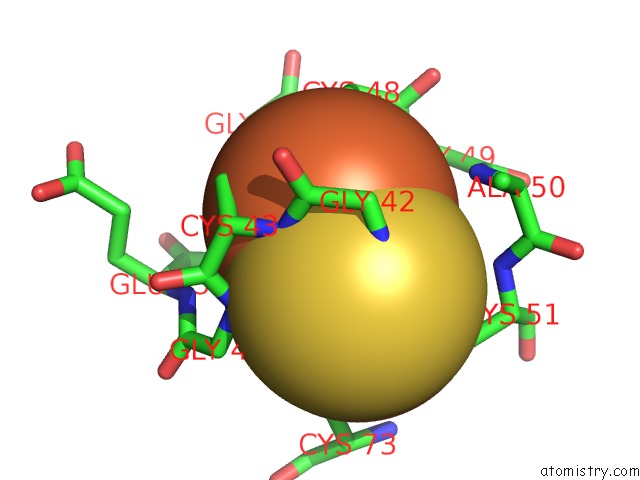
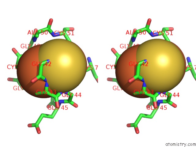
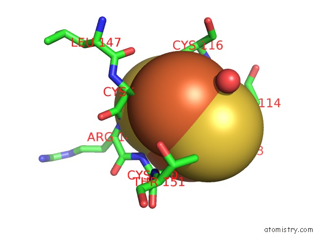
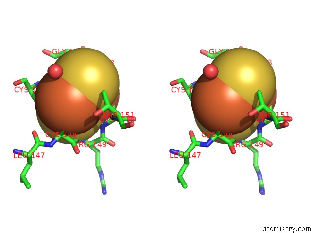
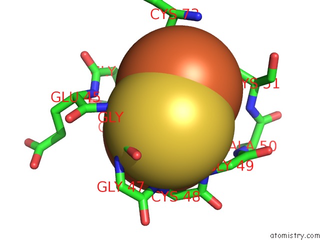
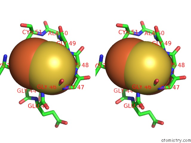
Iron binding site 2 out of 8 in 3unc
Go back to
Iron binding site 2 out
of 8 in the Crystal Structure of Bovine Milk Xanthine Dehydrogenase to 1.65A Resolution
Mono view
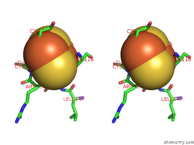
Stereo pair view
Mono view
Stereo pair view
A full contact list of Iron with other atoms in the Fe binding
site number 2 of Crystal Structure of Bovine Milk Xanthine Dehydrogenase to 1.65A Resolution within 5.0Å range:
|
Iron binding site 3 out of 8 in 3unc
Go back to
Iron binding site 3 out
of 8 in the Crystal Structure of Bovine Milk Xanthine Dehydrogenase to 1.65A Resolution
Mono view
Stereo pair view
Mono view
Stereo pair view
A full contact list of Iron with other atoms in the Fe binding
site number 3 of Crystal Structure of Bovine Milk Xanthine Dehydrogenase to 1.65A Resolution within 5.0Å range:
|
Iron binding site 4 out of 8 in 3unc
Go back to
Iron binding site 4 out
of 8 in the Crystal Structure of Bovine Milk Xanthine Dehydrogenase to 1.65A Resolution
Mono view
Stereo pair view
Mono view
Stereo pair view
A full contact list of Iron with other atoms in the Fe binding
site number 4 of Crystal Structure of Bovine Milk Xanthine Dehydrogenase to 1.65A Resolution within 5.0Å range:
|
Iron binding site 5 out of 8 in 3unc
Go back to
Iron binding site 5 out
of 8 in the Crystal Structure of Bovine Milk Xanthine Dehydrogenase to 1.65A Resolution
Mono view
Stereo pair view
Mono view
Stereo pair view
A full contact list of Iron with other atoms in the Fe binding
site number 5 of Crystal Structure of Bovine Milk Xanthine Dehydrogenase to 1.65A Resolution within 5.0Å range:
|
Iron binding site 6 out of 8 in 3unc
Go back to
Iron binding site 6 out
of 8 in the Crystal Structure of Bovine Milk Xanthine Dehydrogenase to 1.65A Resolution
Mono view
Stereo pair view
Mono view
Stereo pair view
A full contact list of Iron with other atoms in the Fe binding
site number 6 of Crystal Structure of Bovine Milk Xanthine Dehydrogenase to 1.65A Resolution within 5.0Å range:
|
Iron binding site 7 out of 8 in 3unc
Go back to
Iron binding site 7 out
of 8 in the Crystal Structure of Bovine Milk Xanthine Dehydrogenase to 1.65A Resolution
Mono view
Stereo pair view
Mono view
Stereo pair view
A full contact list of Iron with other atoms in the Fe binding
site number 7 of Crystal Structure of Bovine Milk Xanthine Dehydrogenase to 1.65A Resolution within 5.0Å range:
|
Iron binding site 8 out of 8 in 3unc
Go back to
Iron binding site 8 out
of 8 in the Crystal Structure of Bovine Milk Xanthine Dehydrogenase to 1.65A Resolution
Mono view
Stereo pair view
Mono view
Stereo pair view
A full contact list of Iron with other atoms in the Fe binding
site number 8 of Crystal Structure of Bovine Milk Xanthine Dehydrogenase to 1.65A Resolution within 5.0Å range:
|
Reference:
H.Ishikita,
B.T.Eger,
K.Okamoto,
T.Nishino,
E.F.Pai.
Protein Conformational Gating of Enzymatic Activity in Xanthine Oxidoreductase. J.Am.Chem.Soc. V. 134 999 2012.
ISSN: ISSN 0002-7863
PubMed: 22145797
DOI: 10.1021/JA207173P
Page generated: Sun Aug 4 21:16:15 2024
ISSN: ISSN 0002-7863
PubMed: 22145797
DOI: 10.1021/JA207173P
Last articles
Zn in 9J0NZn in 9J0O
Zn in 9J0P
Zn in 9FJX
Zn in 9EKB
Zn in 9C0F
Zn in 9CAH
Zn in 9CH0
Zn in 9CH3
Zn in 9CH1