Iron »
PDB 3uhg-3v2z »
3use »
Iron in PDB 3use: Crystal Structure of E. Coli Hydrogenase-1 in Its As-Isolated Form
Enzymatic activity of Crystal Structure of E. Coli Hydrogenase-1 in Its As-Isolated Form
All present enzymatic activity of Crystal Structure of E. Coli Hydrogenase-1 in Its As-Isolated Form:
1.12.99.6;
1.12.99.6;
Protein crystallography data
The structure of Crystal Structure of E. Coli Hydrogenase-1 in Its As-Isolated Form, PDB code: 3use
was solved by
A.Volbeda,
J.C.Fontecilla-Camps,
C.Darnault,
with X-Ray Crystallography technique. A brief refinement statistics is given in the table below:
| Resolution Low / High (Å) | 29.82 / 1.67 |
| Space group | P 21 21 21 |
| Cell size a, b, c (Å), α, β, γ (°) | 93.930, 97.790, 183.290, 90.00, 90.00, 90.00 |
| R / Rfree (%) | 12 / 16.8 |
Other elements in 3use:
The structure of Crystal Structure of E. Coli Hydrogenase-1 in Its As-Isolated Form also contains other interesting chemical elements:
| Nickel | (Ni) | 2 atoms |
| Magnesium | (Mg) | 2 atoms |
| Chlorine | (Cl) | 3 atoms |
Iron Binding Sites:
Pages:
>>> Page 1 <<< Page 2, Binding sites: 11 - 20; Page 3, Binding sites: 21 - 30; Page 4, Binding sites: 31 - 32;Binding sites:
The binding sites of Iron atom in the Crystal Structure of E. Coli Hydrogenase-1 in Its As-Isolated Form (pdb code 3use). This binding sites where shown within 5.0 Angstroms radius around Iron atom.In total 32 binding sites of Iron where determined in the Crystal Structure of E. Coli Hydrogenase-1 in Its As-Isolated Form, PDB code: 3use:
Jump to Iron binding site number: 1; 2; 3; 4; 5; 6; 7; 8; 9; 10;
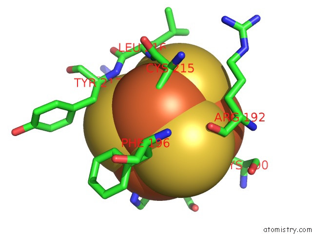
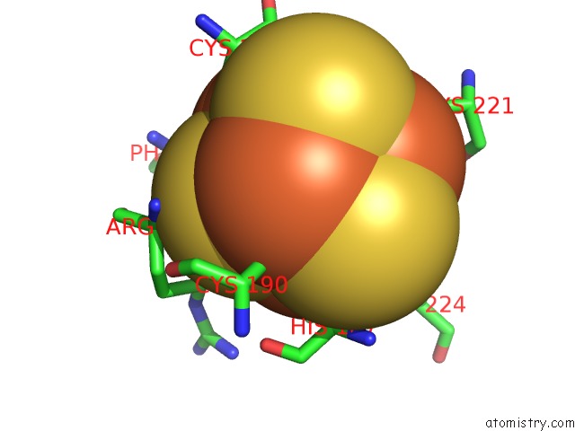
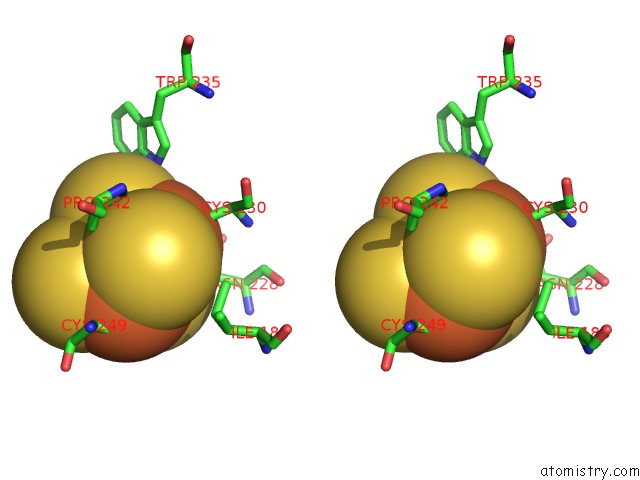
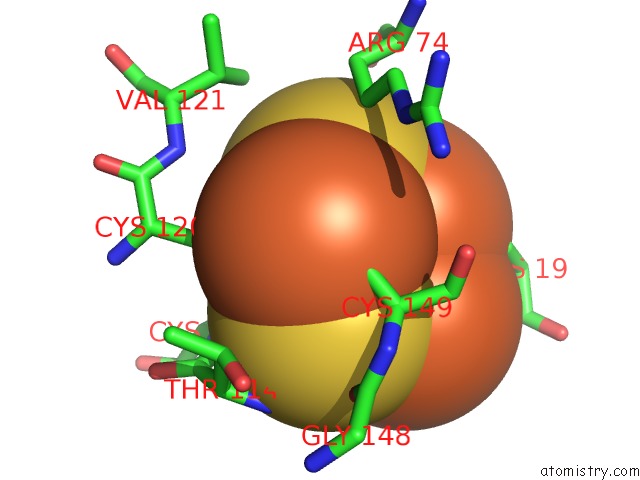
Iron binding site 1 out of 32 in 3use
Go back to
Iron binding site 1 out
of 32 in the Crystal Structure of E. Coli Hydrogenase-1 in Its As-Isolated Form
Mono view
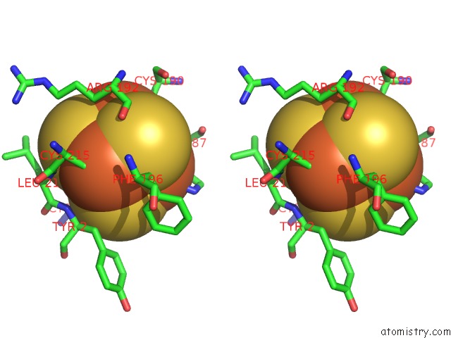
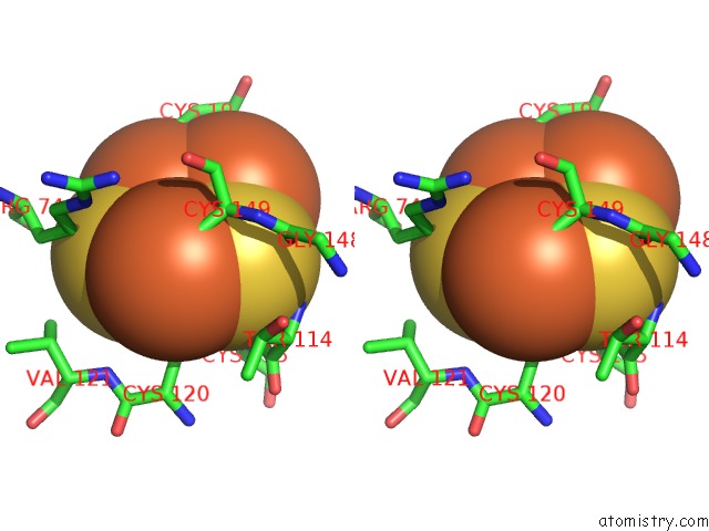
Stereo pair view
Mono view
Stereo pair view
A full contact list of Iron with other atoms in the Fe binding
site number 1 of Crystal Structure of E. Coli Hydrogenase-1 in Its As-Isolated Form within 5.0Å range:
|
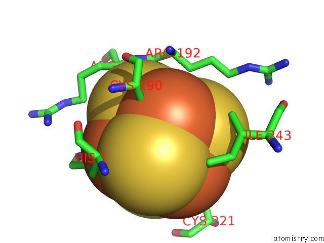
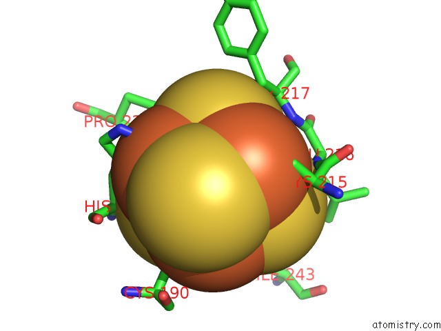
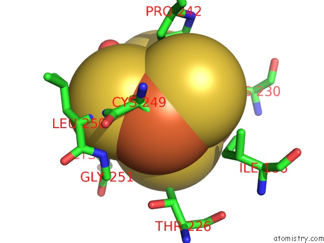
Iron binding site 2 out of 32 in 3use
Go back to
Iron binding site 2 out
of 32 in the Crystal Structure of E. Coli Hydrogenase-1 in Its As-Isolated Form
Mono view
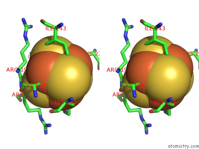
Stereo pair view
Mono view
Stereo pair view
A full contact list of Iron with other atoms in the Fe binding
site number 2 of Crystal Structure of E. Coli Hydrogenase-1 in Its As-Isolated Form within 5.0Å range:
|
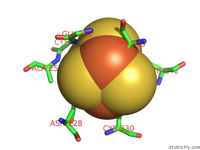
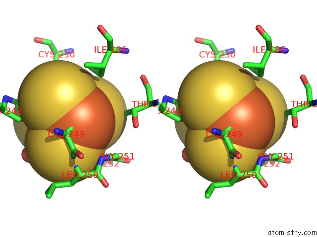
Iron binding site 3 out of 32 in 3use
Go back to
Iron binding site 3 out
of 32 in the Crystal Structure of E. Coli Hydrogenase-1 in Its As-Isolated Form
Mono view

Stereo pair view
Mono view
Stereo pair view
A full contact list of Iron with other atoms in the Fe binding
site number 3 of Crystal Structure of E. Coli Hydrogenase-1 in Its As-Isolated Form within 5.0Å range:
|
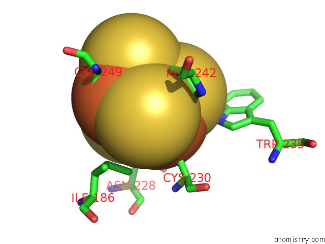
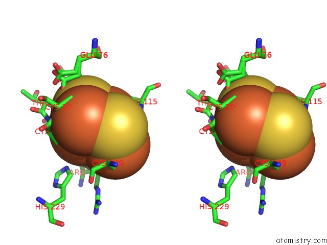
Iron binding site 4 out of 32 in 3use
Go back to
Iron binding site 4 out
of 32 in the Crystal Structure of E. Coli Hydrogenase-1 in Its As-Isolated Form
Mono view

Stereo pair view
Mono view
Stereo pair view
A full contact list of Iron with other atoms in the Fe binding
site number 4 of Crystal Structure of E. Coli Hydrogenase-1 in Its As-Isolated Form within 5.0Å range:
|
Iron binding site 5 out of 32 in 3use
Go back to
Iron binding site 5 out
of 32 in the Crystal Structure of E. Coli Hydrogenase-1 in Its As-Isolated Form
Mono view

Stereo pair view
Mono view
Stereo pair view
A full contact list of Iron with other atoms in the Fe binding
site number 5 of Crystal Structure of E. Coli Hydrogenase-1 in Its As-Isolated Form within 5.0Å range:
|
Iron binding site 6 out of 32 in 3use
Go back to
Iron binding site 6 out
of 32 in the Crystal Structure of E. Coli Hydrogenase-1 in Its As-Isolated Form
Mono view
Stereo pair view
Mono view
Stereo pair view
A full contact list of Iron with other atoms in the Fe binding
site number 6 of Crystal Structure of E. Coli Hydrogenase-1 in Its As-Isolated Form within 5.0Å range:
|
Iron binding site 7 out of 32 in 3use
Go back to
Iron binding site 7 out
of 32 in the Crystal Structure of E. Coli Hydrogenase-1 in Its As-Isolated Form
Mono view
Stereo pair view
Mono view
Stereo pair view
A full contact list of Iron with other atoms in the Fe binding
site number 7 of Crystal Structure of E. Coli Hydrogenase-1 in Its As-Isolated Form within 5.0Å range:
|
Iron binding site 8 out of 32 in 3use
Go back to
Iron binding site 8 out
of 32 in the Crystal Structure of E. Coli Hydrogenase-1 in Its As-Isolated Form

Mono view
Stereo pair view
Mono view
Stereo pair view
A full contact list of Iron with other atoms in the Fe binding
site number 8 of Crystal Structure of E. Coli Hydrogenase-1 in Its As-Isolated Form within 5.0Å range:
|
Iron binding site 9 out of 32 in 3use
Go back to
Iron binding site 9 out
of 32 in the Crystal Structure of E. Coli Hydrogenase-1 in Its As-Isolated Form

Mono view

Stereo pair view
Mono view
Stereo pair view
A full contact list of Iron with other atoms in the Fe binding
site number 9 of Crystal Structure of E. Coli Hydrogenase-1 in Its As-Isolated Form within 5.0Å range:
|
Iron binding site 10 out of 32 in 3use
Go back to
Iron binding site 10 out
of 32 in the Crystal Structure of E. Coli Hydrogenase-1 in Its As-Isolated Form
Mono view
Stereo pair view
Mono view
Stereo pair view
A full contact list of Iron with other atoms in the Fe binding
site number 10 of Crystal Structure of E. Coli Hydrogenase-1 in Its As-Isolated Form within 5.0Å range:
|
Reference:
A.Volbeda,
P.Amara,
C.Darnault,
J.M.Mouesca,
A.Parkin,
M.M.Roessler,
F.A.Armstrong,
J.C.Fontecilla-Camps.
X-Ray Crystallographic and Computational Studies of the O2-Tolerant [Nife]-Hydrogenase 1 From Escherichia Coli. Proc.Natl.Acad.Sci.Usa V. 109 5305 2012.
ISSN: ISSN 0027-8424
PubMed: 22431599
DOI: 10.1073/PNAS.1119806109
Page generated: Sun Aug 4 21:17:12 2024
ISSN: ISSN 0027-8424
PubMed: 22431599
DOI: 10.1073/PNAS.1119806109
Last articles
Zn in 9JYWZn in 9IR4
Zn in 9IR3
Zn in 9GMX
Zn in 9GMW
Zn in 9JEJ
Zn in 9ERF
Zn in 9ERE
Zn in 9EGV
Zn in 9EGW