Atomistry » Iron » PDB 3uhg-3v2z » 3uus
Atomistry »
  Iron »
    PDB 3uhg-3v2z »
      3uus »

Iron in PDB 3uus: Crystal Structure of the Datp Inhibited E. Coli Class Ia Ribonucleotide Reductase Complex

Enzymatic activity of Crystal Structure of the Datp Inhibited E. Coli Class Ia Ribonucleotide Reductase Complex

All present enzymatic activity of Crystal Structure of the Datp Inhibited E. Coli Class Ia Ribonucleotide Reductase Complex:
1.17.4.1;

Protein crystallography data

The structure of Crystal Structure of the Datp Inhibited E. Coli Class Ia Ribonucleotide Reductase Complex, PDB code: 3uus was solved by C.M.Zimanyi, C.L.Drennan, with X-Ray Crystallography technique. A brief refinement statistics is given in the table below:

Resolution Low / High (Å) 50.00 / 5.65
Space group C 1 2 1
Cell size a, b, c (Å), α, β, γ (°) 287.358, 153.457, 169.416, 90.00, 119.91, 90.00
R / Rfree (%) 25.7 / 30.3

Iron Binding Sites:

The binding sites of Iron atom in the Crystal Structure of the Datp Inhibited E. Coli Class Ia Ribonucleotide Reductase Complex (pdb code 3uus). This binding sites where shown within 5.0 Angstroms radius around Iron atom.
In total 8 binding sites of Iron where determined in the Crystal Structure of the Datp Inhibited E. Coli Class Ia Ribonucleotide Reductase Complex, PDB code: 3uus:
Jump to Iron binding site number: 1; 2; 3; 4; 5; 6; 7; 8;

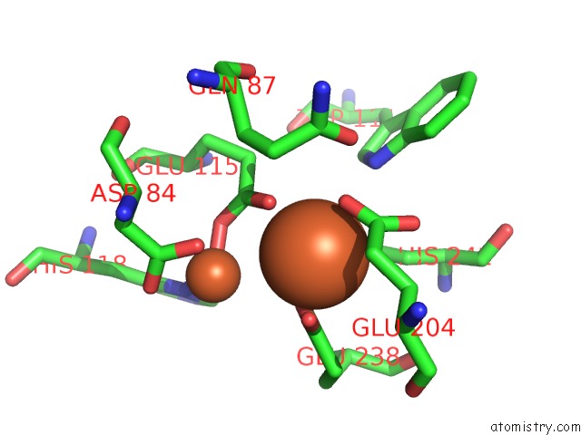
Iron binding site 1 out of 8 in 3uus

Go back to Iron Binding Sites List in 3uus
Iron binding site 1 out of 8 in the Crystal Structure of the Datp Inhibited E. Coli Class Ia Ribonucleotide Reductase Complex


Mono view


Stereo pair view

A full contact list of Iron with other atoms in the Fe binding site number 1 of Crystal Structure of the Datp Inhibited E. Coli Class Ia Ribonucleotide Reductase Complex within 5.0Å range:
probe atom residue distance (Å) B Occ
E:Fe1003

b:0.3
occ:1.00
OE1 E:GLU115 1.7 0.6 1.0
ND1 E:HIS118 2.0 0.1 1.0
OD2 E:ASP84 2.3 0.9 1.0
OD1 E:ASP84 2.3 0.9 1.0
CG E:ASP84 2.5 0.9 1.0
CE1 E:HIS118 2.7 0.1 1.0
CD E:GLU115 2.8 0.6 1.0
OE1 E:GLU238 3.1 0.3 1.0
OE2 E:GLU115 3.2 0.6 1.0
CG E:HIS118 3.2 0.1 1.0
FE E:FE1004 3.4 0.1 1.0
CB E:HIS118 3.8 0.1 1.0
CD E:GLU238 3.8 0.3 1.0
CB E:ASP84 3.9 0.9 1.0
NE2 E:HIS118 3.9 0.1 1.0
OE2 E:GLU238 4.0 0.3 1.0
CG E:GLU115 4.1 0.6 1.0
CG2 E:ILE234 4.1 0.2 1.0
CD2 E:HIS118 4.1 0.1 1.0
CA E:GLU115 4.4 0.6 1.0
CE1 E:HIS241 4.4 0.6 1.0
CZ E:PHE208 4.4 0.4 1.0
ND1 E:HIS241 4.5 0.6 1.0
CE2 E:PHE208 4.5 0.4 1.0
CB E:GLU115 4.6 0.6 1.0
CA E:ASP84 4.8 0.9 1.0
O E:GLU115 4.9 0.6 1.0
CE1 E:PHE208 5.0 0.4 1.0

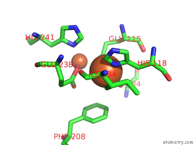
Iron binding site 2 out of 8 in 3uus

Go back to Iron Binding Sites List in 3uus
Iron binding site 2 out of 8 in the Crystal Structure of the Datp Inhibited E. Coli Class Ia Ribonucleotide Reductase Complex


Mono view


Stereo pair view

A full contact list of Iron with other atoms in the Fe binding site number 2 of Crystal Structure of the Datp Inhibited E. Coli Class Ia Ribonucleotide Reductase Complex within 5.0Å range:
probe atom residue distance (Å) B Occ
E:Fe1004

b:0.1
occ:1.00
OE1 E:GLU238 1.8 0.3 1.0
ND1 E:HIS241 2.2 0.6 1.0
OE1 E:GLU204 2.3 0.8 1.0
OE2 E:GLU115 2.3 0.6 1.0
CD E:GLU238 2.5 0.3 1.0
OE2 E:GLU238 2.7 0.3 1.0
CD E:GLU204 2.8 0.8 1.0
CD E:GLU115 2.9 0.6 1.0
CE1 E:HIS241 3.1 0.6 1.0
CG E:HIS241 3.2 0.6 1.0
OE1 E:GLU115 3.3 0.6 1.0
FE E:FE1003 3.4 0.3 1.0
CG E:GLU204 3.4 0.8 1.0
OE2 E:GLU204 3.4 0.8 1.0
CB E:HIS241 3.5 0.6 1.0
CG E:GLU238 3.8 0.3 1.0
NE1 E:TRP111 3.8 1.0 1.0
CG E:GLU115 4.0 0.6 1.0
OD1 E:ASP84 4.1 0.9 1.0
CA E:GLU238 4.2 0.3 1.0
NE2 E:HIS241 4.3 0.6 1.0
CB E:GLU204 4.3 0.8 1.0
CD2 E:HIS241 4.3 0.6 1.0
CB E:GLU238 4.4 0.3 1.0
CD1 E:TRP111 4.5 1.0 1.0
CE1 E:HIS118 4.8 0.1 1.0
OE1 E:GLN87 4.8 0.9 1.0
ND1 E:HIS118 4.9 0.1 1.0
CE2 E:TRP111 5.0 1.0 1.0

Iron binding site 3 out of 8 in 3uus

Go back to Iron Binding Sites List in 3uus
Iron binding site 3 out of 8 in the Crystal Structure of the Datp Inhibited E. Coli Class Ia Ribonucleotide Reductase Complex


Mono view


Stereo pair view

A full contact list of Iron with other atoms in the Fe binding site number 3 of Crystal Structure of the Datp Inhibited E. Coli Class Ia Ribonucleotide Reductase Complex within 5.0Å range:
probe atom residue distance (Å) B Occ
F:Fe1001

b:94.4
occ:1.00
OE1 F:GLU115 1.7 0.7 1.0
ND1 F:HIS118 2.0 0.1 1.0
OD2 F:ASP84 2.2 0.8 1.0
OD1 F:ASP84 2.3 0.8 1.0
CG F:ASP84 2.5 0.8 1.0
CD F:GLU115 2.7 0.7 1.0
CE1 F:HIS118 2.7 0.1 1.0
OE2 F:GLU115 3.1 0.7 1.0
OE1 F:GLU238 3.1 0.8 1.0
CG F:HIS118 3.2 0.1 1.0
FE F:FE1002 3.4 1.0 1.0
CB F:HIS118 3.8 0.1 1.0
CD F:GLU238 3.8 0.8 1.0
CB F:ASP84 3.8 0.8 1.0
NE2 F:HIS118 3.9 0.1 1.0
OE2 F:GLU238 4.0 0.8 1.0
CG F:GLU115 4.1 0.7 1.0
CG2 F:ILE234 4.1 0.3 1.0
CD2 F:HIS118 4.2 0.1 1.0
CA F:GLU115 4.3 0.7 1.0
CZ F:PHE208 4.4 0.0 1.0
CE1 F:HIS241 4.4 0.7 1.0
CE2 F:PHE208 4.5 0.0 1.0
ND1 F:HIS241 4.5 0.7 1.0
CB F:GLU115 4.5 0.7 1.0
CA F:ASP84 4.7 0.8 1.0
O F:GLU115 4.9 0.7 1.0
CE1 F:PHE208 5.0 0.0 1.0

Iron binding site 4 out of 8 in 3uus

Go back to Iron Binding Sites List in 3uus
Iron binding site 4 out of 8 in the Crystal Structure of the Datp Inhibited E. Coli Class Ia Ribonucleotide Reductase Complex


Mono view


Stereo pair view

A full contact list of Iron with other atoms in the Fe binding site number 4 of Crystal Structure of the Datp Inhibited E. Coli Class Ia Ribonucleotide Reductase Complex within 5.0Å range:
probe atom residue distance (Å) B Occ
F:Fe1002

b:1.0
occ:1.00
OE1 F:GLU238 1.8 0.8 1.0
ND1 F:HIS241 2.2 0.7 1.0
OE2 F:GLU115 2.3 0.7 1.0
OE1 F:GLU204 2.3 0.9 1.0
CD F:GLU238 2.5 0.8 1.0
OE2 F:GLU238 2.8 0.8 1.0
CD F:GLU204 2.8 0.9 1.0
CD F:GLU115 2.9 0.7 1.0
CE1 F:HIS241 3.1 0.7 1.0
CG F:HIS241 3.2 0.7 1.0
OE1 F:GLU115 3.2 0.7 1.0
FE F:FE1001 3.4 94.4 1.0
OE2 F:GLU204 3.4 0.9 1.0
CG F:GLU204 3.5 0.9 1.0
CB F:HIS241 3.5 0.7 1.0
NE1 F:TRP111 3.8 0.0 1.0
CG F:GLU238 3.9 0.8 1.0
CG F:GLU115 3.9 0.7 1.0
OD1 F:ASP84 4.1 0.8 1.0
CA F:GLU238 4.2 0.8 1.0
NE2 F:HIS241 4.2 0.7 1.0
CD2 F:HIS241 4.3 0.7 1.0
CB F:GLU204 4.3 0.9 1.0
CB F:GLU238 4.4 0.8 1.0
CD1 F:TRP111 4.4 0.0 1.0
CE1 F:HIS118 4.7 0.1 1.0
OE1 F:GLN87 4.8 0.5 1.0
ND1 F:HIS118 4.9 0.1 1.0
CE2 F:TRP111 4.9 0.0 1.0

Iron binding site 5 out of 8 in 3uus

Go back to Iron Binding Sites List in 3uus
Iron binding site 5 out of 8 in the Crystal Structure of the Datp Inhibited E. Coli Class Ia Ribonucleotide Reductase Complex


Mono view


Stereo pair view

A full contact list of Iron with other atoms in the Fe binding site number 5 of Crystal Structure of the Datp Inhibited E. Coli Class Ia Ribonucleotide Reductase Complex within 5.0Å range:
probe atom residue distance (Å) B Occ
G:Fe1003

b:89.7
occ:1.00
OE1 G:GLU115 1.7 0.8 1.0
ND1 G:HIS118 2.0 0.0 1.0
OD1 G:ASP84 2.3 0.0 1.0
OD2 G:ASP84 2.3 0.0 1.0
CG G:ASP84 2.5 0.0 1.0
CE1 G:HIS118 2.7 0.0 1.0
CD G:GLU115 2.7 0.8 1.0
OE1 G:GLU238 3.0 0.8 1.0
OE2 G:GLU115 3.1 0.8 1.0
CG G:HIS118 3.2 0.0 1.0
FE G:FE1004 3.4 0.8 1.0
CD G:GLU238 3.7 0.8 1.0
CB G:HIS118 3.8 0.0 1.0
NE2 G:HIS118 3.9 0.0 1.0
CB G:ASP84 3.9 0.0 1.0
OE2 G:GLU238 3.9 0.8 1.0
CG G:GLU115 4.1 0.8 1.0
CG2 G:ILE234 4.1 0.5 1.0
CD2 G:HIS118 4.2 0.0 1.0
CE1 G:HIS241 4.3 0.4 1.0
CA G:GLU115 4.4 0.8 1.0
ND1 G:HIS241 4.4 0.4 1.0
CZ G:PHE208 4.4 0.8 1.0
CE2 G:PHE208 4.5 0.8 1.0
CB G:GLU115 4.6 0.8 1.0
CA G:ASP84 4.8 0.0 1.0
O G:GLU115 4.9 0.8 1.0
CG G:GLU238 5.0 0.8 1.0
CE1 G:PHE208 5.0 0.8 1.0

Iron binding site 6 out of 8 in 3uus

Go back to Iron Binding Sites List in 3uus
Iron binding site 6 out of 8 in the Crystal Structure of the Datp Inhibited E. Coli Class Ia Ribonucleotide Reductase Complex


Mono view


Stereo pair view

A full contact list of Iron with other atoms in the Fe binding site number 6 of Crystal Structure of the Datp Inhibited E. Coli Class Ia Ribonucleotide Reductase Complex within 5.0Å range:
probe atom residue distance (Å) B Occ
G:Fe1004

b:0.8
occ:1.00
OE1 G:GLU238 1.8 0.8 1.0
ND1 G:HIS241 2.2 0.4 1.0
OE1 G:GLU204 2.3 0.4 1.0
OE2 G:GLU115 2.4 0.8 1.0
CD G:GLU238 2.5 0.8 1.0
CD G:GLU204 2.7 0.4 1.0
OE2 G:GLU238 2.7 0.8 1.0
CD G:GLU115 3.0 0.8 1.0
CE1 G:HIS241 3.1 0.4 1.0
CG G:HIS241 3.2 0.4 1.0
OE2 G:GLU204 3.3 0.4 1.0
OE1 G:GLU115 3.3 0.8 1.0
CG G:GLU204 3.4 0.4 1.0
FE G:FE1003 3.4 89.7 1.0
CB G:HIS241 3.5 0.4 1.0
CG G:GLU238 3.8 0.8 1.0
NE1 G:TRP111 3.8 0.8 1.0
CG G:GLU115 4.0 0.8 1.0
CA G:GLU238 4.2 0.8 1.0
OD1 G:ASP84 4.2 0.0 1.0
CB G:GLU204 4.2 0.4 1.0
NE2 G:HIS241 4.3 0.4 1.0
CD2 G:HIS241 4.3 0.4 1.0
CB G:GLU238 4.4 0.8 1.0
CD1 G:TRP111 4.4 0.8 1.0
OE1 G:GLN87 4.8 0.6 1.0
CE1 G:HIS118 4.8 0.0 1.0
CE2 G:TRP111 4.9 0.8 1.0
O G:GLU238 5.0 0.8 1.0
ND1 G:HIS118 5.0 0.0 1.0
CA G:HIS241 5.0 0.4 1.0

Iron binding site 7 out of 8 in 3uus

Go back to Iron Binding Sites List in 3uus
Iron binding site 7 out of 8 in the Crystal Structure of the Datp Inhibited E. Coli Class Ia Ribonucleotide Reductase Complex


Mono view


Stereo pair view

A full contact list of Iron with other atoms in the Fe binding site number 7 of Crystal Structure of the Datp Inhibited E. Coli Class Ia Ribonucleotide Reductase Complex within 5.0Å range:
probe atom residue distance (Å) B Occ
H:Fe1001

b:0.9
occ:1.00
OE1 H:GLU115 1.7 0.8 1.0
ND1 H:HIS118 2.0 0.0 1.0
OD2 H:ASP84 2.2 0.6 1.0
OD1 H:ASP84 2.3 0.6 1.0
CG H:ASP84 2.4 0.6 1.0
CD H:GLU115 2.7 0.8 1.0
CE1 H:HIS118 2.8 0.0 1.0
OE2 H:GLU115 3.1 0.8 1.0
OE1 H:GLU238 3.2 0.6 1.0
CG H:HIS118 3.2 0.0 1.0
FE H:FE1002 3.4 0.5 1.0
CB H:HIS118 3.8 0.0 1.0
CB H:ASP84 3.8 0.6 1.0
CD H:GLU238 3.9 0.6 1.0
NE2 H:HIS118 3.9 0.0 1.0
OE2 H:GLU238 4.0 0.6 1.0
CG H:GLU115 4.1 0.8 1.0
CG2 H:ILE234 4.2 0.2 1.0
CD2 H:HIS118 4.2 0.0 1.0
CA H:GLU115 4.3 0.8 1.0
CZ H:PHE208 4.3 0.9 1.0
CE1 H:HIS241 4.5 0.2 1.0
CE2 H:PHE208 4.5 0.9 1.0
ND1 H:HIS241 4.5 0.2 1.0
CB H:GLU115 4.5 0.8 1.0
CA H:ASP84 4.7 0.6 1.0
O H:GLU115 4.8 0.8 1.0
CE1 H:PHE208 4.9 0.9 1.0

Iron binding site 8 out of 8 in 3uus

Go back to Iron Binding Sites List in 3uus
Iron binding site 8 out of 8 in the Crystal Structure of the Datp Inhibited E. Coli Class Ia Ribonucleotide Reductase Complex


Mono view


Stereo pair view

A full contact list of Iron with other atoms in the Fe binding site number 8 of Crystal Structure of the Datp Inhibited E. Coli Class Ia Ribonucleotide Reductase Complex within 5.0Å range:
probe atom residue distance (Å) B Occ
H:Fe1002

b:0.5
occ:1.00
OE1 H:GLU238 1.8 0.6 1.0
ND1 H:HIS241 2.2 0.2 1.0
OE2 H:GLU115 2.3 0.8 1.0
OE1 H:GLU204 2.3 0.3 1.0
CD H:GLU238 2.5 0.6 1.0
CD H:GLU204 2.8 0.3 1.0
CD H:GLU115 2.8 0.8 1.0
OE2 H:GLU238 2.8 0.6 1.0
CE1 H:HIS241 3.1 0.2 1.0
OE1 H:GLU115 3.2 0.8 1.0
CG H:HIS241 3.2 0.2 1.0
FE H:FE1001 3.4 0.9 1.0
OE2 H:GLU204 3.4 0.3 1.0
CG H:GLU204 3.5 0.3 1.0
CB H:HIS241 3.6 0.2 1.0
NE1 H:TRP111 3.8 0.4 1.0
CG H:GLU238 3.9 0.6 1.0
CG H:GLU115 3.9 0.8 1.0
OD1 H:ASP84 4.1 0.6 1.0
NE2 H:HIS241 4.2 0.2 1.0
CA H:GLU238 4.2 0.6 1.0
CD2 H:HIS241 4.3 0.2 1.0
CB H:GLU204 4.4 0.3 1.0
CB H:GLU238 4.4 0.6 1.0
CD1 H:TRP111 4.4 0.4 1.0
CE1 H:HIS118 4.7 0.0 1.0
OE1 H:GLN87 4.8 0.5 1.0
ND1 H:HIS118 4.9 0.0 1.0
CE2 H:TRP111 4.9 0.4 1.0
CB H:GLN87 5.0 0.5 1.0

Reference:

N.Ando, E.J.Brignole, C.M.Zimanyi, M.A.Funk, K.Yokoyama, F.J.Asturias, J.Stubbe, C.L.Drennan. Structural Interconversions Modulate Activity of Escherichia Coli Ribonucleotide Reductase. Proc.Natl.Acad.Sci.Usa V. 108 21046 2011.
ISSN: ISSN 0027-8424
PubMed: 22160671
DOI: 10.1073/PNAS.1112715108
Page generated: Sun Aug 4 21:25:58 2024

Last articles

Zn in 9J0N
Zn in 9J0O
Zn in 9J0P
Zn in 9FJX
Zn in 9EKB
Zn in 9C0F
Zn in 9CAH
Zn in 9CH0
Zn in 9CH3
Zn in 9CH1
© Copyright 2008-2020 by atomistry.com
Home   |    Site Map   |    Copyright   |    Contact us   |    Privacy