Iron »
PDB 3vkt-3vti »
3vm9 »
Iron in PDB 3vm9: Dimeric Horse Myoglobin
Protein crystallography data
The structure of Dimeric Horse Myoglobin, PDB code: 3vm9
was solved by
S.Nagao,
H.Osuka,
T.Yamada,
T.Uni,
Y.Shomura,
K.Imai,
Y.Higuchi,
S.Hirota,
with X-Ray Crystallography technique. A brief refinement statistics is given in the table below:
| Resolution Low / High (Å) | 20.00 / 1.05 |
| Space group | P 21 21 21 |
| Cell size a, b, c (Å), α, β, γ (°) | 57.340, 62.523, 83.354, 90.00, 90.00, 90.00 |
| R / Rfree (%) | 12.8 / 16.8 |
Iron Binding Sites:
The binding sites of Iron atom in the Dimeric Horse Myoglobin
(pdb code 3vm9). This binding sites where shown within
5.0 Angstroms radius around Iron atom.
In total 2 binding sites of Iron where determined in the Dimeric Horse Myoglobin, PDB code: 3vm9:
Jump to Iron binding site number: 1; 2;
In total 2 binding sites of Iron where determined in the Dimeric Horse Myoglobin, PDB code: 3vm9:
Jump to Iron binding site number: 1; 2;
Iron binding site 1 out of 2 in 3vm9
Go back to
Iron binding site 1 out
of 2 in the Dimeric Horse Myoglobin
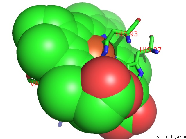
Mono view

Stereo pair view
Mono view
Stereo pair view
A full contact list of Iron with other atoms in the Fe binding
site number 1 of Dimeric Horse Myoglobin within 5.0Å range:
|
Iron binding site 2 out of 2 in 3vm9
Go back to
Iron binding site 2 out
of 2 in the Dimeric Horse Myoglobin
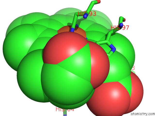
Mono view
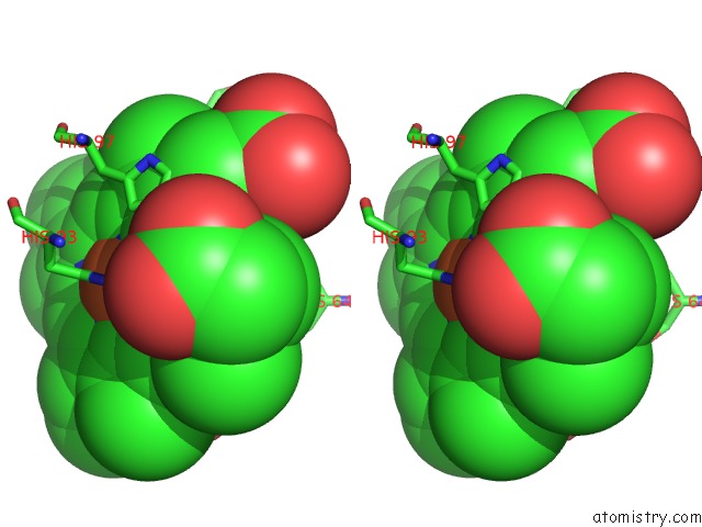
Stereo pair view
Mono view
Stereo pair view
A full contact list of Iron with other atoms in the Fe binding
site number 2 of Dimeric Horse Myoglobin within 5.0Å range:
|
Reference:
S.Nagao,
H.Osuka,
T.Yamada,
T.Uni,
Y.Shomura,
K.Imai,
Y.Higuchi,
S.Hirota.
Structural and Oxygen Binding Properties of Dimeric Horse Myoglobin Dalton Trans V. 41 11378 2012.
ISSN: ISSN 1477-9226
PubMed: 22885714
DOI: 10.1039/C2DT30893B
Page generated: Sun Aug 4 22:03:01 2024
ISSN: ISSN 1477-9226
PubMed: 22885714
DOI: 10.1039/C2DT30893B
Last articles
Zn in 9J0NZn in 9J0O
Zn in 9J0P
Zn in 9FJX
Zn in 9EKB
Zn in 9C0F
Zn in 9CAH
Zn in 9CH0
Zn in 9CH3
Zn in 9CH1