Iron »
PDB 3vkt-3vti »
3vol »
Iron in PDB 3vol: X-Ray Crystal Structure of Pas-Hamp AER2 in the Cn-Bound Form
Protein crystallography data
The structure of X-Ray Crystal Structure of Pas-Hamp AER2 in the Cn-Bound Form, PDB code: 3vol
was solved by
H.Sawai,
H.Sugimoto,
Y.Shiro,
S.Aono,
with X-Ray Crystallography technique. A brief refinement statistics is given in the table below:
| Resolution Low / High (Å) | 29.90 / 2.40 |
| Space group | P 65 2 2 |
| Cell size a, b, c (Å), α, β, γ (°) | 83.020, 83.020, 107.760, 90.00, 90.00, 120.00 |
| R / Rfree (%) | 22.3 / 26.8 |
Iron Binding Sites:
The binding sites of Iron atom in the X-Ray Crystal Structure of Pas-Hamp AER2 in the Cn-Bound Form
(pdb code 3vol). This binding sites where shown within
5.0 Angstroms radius around Iron atom.
In total only one binding site of Iron was determined in the X-Ray Crystal Structure of Pas-Hamp AER2 in the Cn-Bound Form, PDB code: 3vol:
In total only one binding site of Iron was determined in the X-Ray Crystal Structure of Pas-Hamp AER2 in the Cn-Bound Form, PDB code: 3vol:
Iron binding site 1 out of 1 in 3vol
Go back to
Iron binding site 1 out
of 1 in the X-Ray Crystal Structure of Pas-Hamp AER2 in the Cn-Bound Form
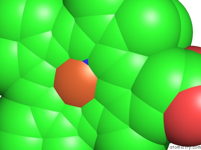
Mono view
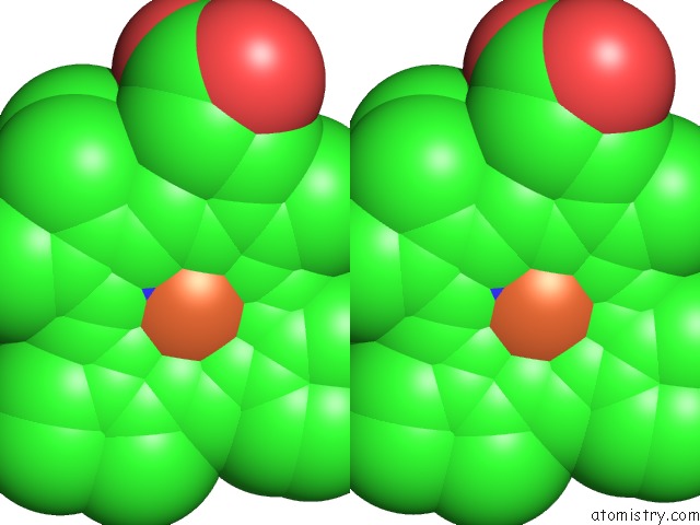
Stereo pair view
Mono view
Stereo pair view
A full contact list of Iron with other atoms in the Fe binding
site number 1 of X-Ray Crystal Structure of Pas-Hamp AER2 in the Cn-Bound Form within 5.0Å range:
|
Reference:
H.Sawai,
H.Sugimoto,
Y.Shiro,
H.Ishikawa,
Y.Mizutani,
S.Aono.
Structural Basis For Oxygen Sensing and Signal Transduction of the Heme-Based Sensor Protein AER2 From Pseudomonas Aeruginosa Chem.Commun.(Camb.) V. 48 6523 2012.
ISSN: ISSN 1359-7345
PubMed: 22622145
DOI: 10.1039/C2CC32549G
Page generated: Sun Aug 4 22:07:07 2024
ISSN: ISSN 1359-7345
PubMed: 22622145
DOI: 10.1039/C2CC32549G
Last articles
Zn in 9J0NZn in 9J0O
Zn in 9J0P
Zn in 9FJX
Zn in 9EKB
Zn in 9C0F
Zn in 9CAH
Zn in 9CH0
Zn in 9CH3
Zn in 9CH1