Iron »
PDB 3whm-3x16 »
3wku »
Iron in PDB 3wku: Crystal Structure of the Anaerobic Desb-Gallate Complex
Enzymatic activity of Crystal Structure of the Anaerobic Desb-Gallate Complex
All present enzymatic activity of Crystal Structure of the Anaerobic Desb-Gallate Complex:
1.13.11.58;
1.13.11.58;
Protein crystallography data
The structure of Crystal Structure of the Anaerobic Desb-Gallate Complex, PDB code: 3wku
was solved by
K.Sugimoto,
M.Senda,
D.Kasai,
M.Fukuda,
E.Masai,
T.Senda,
with X-Ray Crystallography technique. A brief refinement statistics is given in the table below:
| Resolution Low / High (Å) | 20.00 / 2.70 |
| Space group | P 1 21 1 |
| Cell size a, b, c (Å), α, β, γ (°) | 57.435, 60.765, 117.986, 90.00, 98.58, 90.00 |
| R / Rfree (%) | 23.3 / 31.4 |
Iron Binding Sites:
The binding sites of Iron atom in the Crystal Structure of the Anaerobic Desb-Gallate Complex
(pdb code 3wku). This binding sites where shown within
5.0 Angstroms radius around Iron atom.
In total 3 binding sites of Iron where determined in the Crystal Structure of the Anaerobic Desb-Gallate Complex, PDB code: 3wku:
Jump to Iron binding site number: 1; 2; 3;
In total 3 binding sites of Iron where determined in the Crystal Structure of the Anaerobic Desb-Gallate Complex, PDB code: 3wku:
Jump to Iron binding site number: 1; 2; 3;
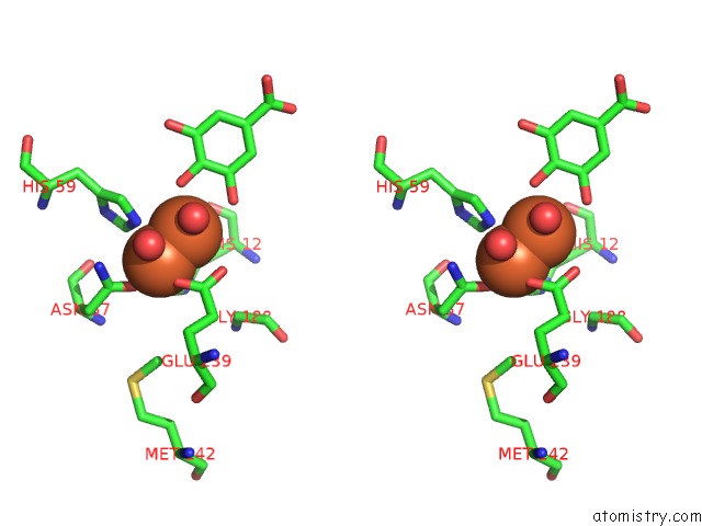
Iron binding site 1 out of 3 in 3wku
Go back to
Iron binding site 1 out
of 3 in the Crystal Structure of the Anaerobic Desb-Gallate Complex

Mono view

Stereo pair view
Mono view
Stereo pair view
A full contact list of Iron with other atoms in the Fe binding
site number 1 of Crystal Structure of the Anaerobic Desb-Gallate Complex within 5.0Å range:
|
Iron binding site 2 out of 3 in 3wku
Go back to
Iron binding site 2 out
of 3 in the Crystal Structure of the Anaerobic Desb-Gallate Complex

Mono view
Stereo pair view
Mono view
Stereo pair view
A full contact list of Iron with other atoms in the Fe binding
site number 2 of Crystal Structure of the Anaerobic Desb-Gallate Complex within 5.0Å range:
|
Iron binding site 3 out of 3 in 3wku
Go back to
Iron binding site 3 out
of 3 in the Crystal Structure of the Anaerobic Desb-Gallate Complex

Mono view

Stereo pair view
Mono view
Stereo pair view
A full contact list of Iron with other atoms in the Fe binding
site number 3 of Crystal Structure of the Anaerobic Desb-Gallate Complex within 5.0Å range:
|
Reference:
K.Sugimoto,
M.Senda,
D.Kasai,
M.Fukuda,
E.Masai,
T.Senda.
Molecular Mechanism of Strict Substrate Specificity of An Extradiol Dioxygenase, Desb, Derived From Sphingobium Sp. Syk-6 Plos One V. 9 92249 2014.
ISSN: ESSN 1932-6203
PubMed: 24657997
DOI: 10.1371/JOURNAL.PONE.0092249
Page generated: Sun Aug 4 22:43:01 2024
ISSN: ESSN 1932-6203
PubMed: 24657997
DOI: 10.1371/JOURNAL.PONE.0092249
Last articles
Zn in 9J0NZn in 9J0O
Zn in 9J0P
Zn in 9FJX
Zn in 9EKB
Zn in 9C0F
Zn in 9CAH
Zn in 9CH0
Zn in 9CH3
Zn in 9CH1