Iron »
PDB 3whm-3x16 »
3wve »
Iron in PDB 3wve: Crystal Structure of Nitrile Hydratase Mutant BR56K Complexed with Trimethylacetonitrile, Before Photo-Activation
Enzymatic activity of Crystal Structure of Nitrile Hydratase Mutant BR56K Complexed with Trimethylacetonitrile, Before Photo-Activation
All present enzymatic activity of Crystal Structure of Nitrile Hydratase Mutant BR56K Complexed with Trimethylacetonitrile, Before Photo-Activation:
4.2.1.84;
4.2.1.84;
Protein crystallography data
The structure of Crystal Structure of Nitrile Hydratase Mutant BR56K Complexed with Trimethylacetonitrile, Before Photo-Activation, PDB code: 3wve
was solved by
Y.Yamanaka,
K.Hashimoto,
K.Noguchi,
M.Yohda,
M.Odaka,
with X-Ray Crystallography technique. A brief refinement statistics is given in the table below:
| Resolution Low / High (Å) | 29.91 / 1.57 |
| Space group | C 1 2 1 |
| Cell size a, b, c (Å), α, β, γ (°) | 113.742, 59.829, 81.464, 90.00, 125.21, 90.00 |
| R / Rfree (%) | 13 / 17.1 |
Other elements in 3wve:
The structure of Crystal Structure of Nitrile Hydratase Mutant BR56K Complexed with Trimethylacetonitrile, Before Photo-Activation also contains other interesting chemical elements:
| Magnesium | (Mg) | 2 atoms |
| Chlorine | (Cl) | 4 atoms |
Iron Binding Sites:
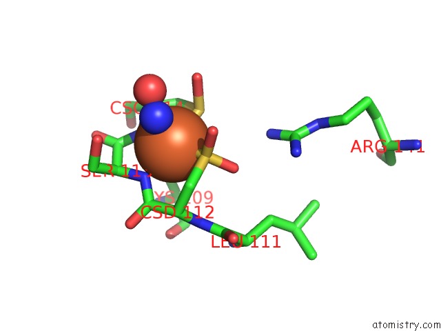
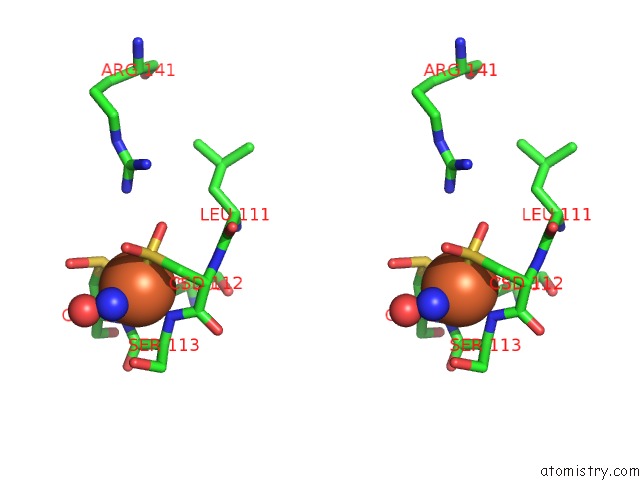
The binding sites of Iron atom in the Crystal Structure of Nitrile Hydratase Mutant BR56K Complexed with Trimethylacetonitrile, Before Photo-Activation
(pdb code 3wve). This binding sites where shown within
5.0 Angstroms radius around Iron atom.
In total only one binding site of Iron was determined in the Crystal Structure of Nitrile Hydratase Mutant BR56K Complexed with Trimethylacetonitrile, Before Photo-Activation, PDB code: 3wve:
In total only one binding site of Iron was determined in the Crystal Structure of Nitrile Hydratase Mutant BR56K Complexed with Trimethylacetonitrile, Before Photo-Activation, PDB code: 3wve:
Iron binding site 1 out of 1 in 3wve
Go back to
Iron binding site 1 out
of 1 in the Crystal Structure of Nitrile Hydratase Mutant BR56K Complexed with Trimethylacetonitrile, Before Photo-Activation
Mono view
Stereo pair view
Mono view
Stereo pair view
A full contact list of Iron with other atoms in the Fe binding
site number 1 of Crystal Structure of Nitrile Hydratase Mutant BR56K Complexed with Trimethylacetonitrile, Before Photo-Activation within 5.0Å range:
|
Reference:
Y.Yamanaka,
Y.Kato,
K.Hashimoto,
K.Iida,
K.Nagasawa,
H.Nakayama,
N.Dohmae,
K.Noguchi,
T.Noguchi,
M.Yohda,
M.Odaka.
Time-Resolved Crystallography of the Reaction Intermediate of Nitrile Hydratase: Revealing A Role For the Cysteinesulfenic Acid Ligand As A Catalytic Nucleophile. Angew.Chem.Int.Ed.Engl. V. 54 10763 2015.
ISSN: ISSN 1433-7851
PubMed: 26333053
DOI: 10.1002/ANIE.201502731
Page generated: Sun Aug 4 22:52:40 2024
ISSN: ISSN 1433-7851
PubMed: 26333053
DOI: 10.1002/ANIE.201502731
Last articles
F in 4LTSF in 4LUD
F in 4LUV
F in 4LOQ
F in 4LQG
F in 4LPB
F in 4LOP
F in 4LP0
F in 4LOY
F in 4LMN