Iron »
PDB 3x20-3zjo »
3x2q »
Iron in PDB 3x2q: X-Ray Structure of Cyanide-Bound Bovine Heart Cytochrome C Oxidase in the Fully Oxidized State at 2.0 Angstrom Resolution
Enzymatic activity of X-Ray Structure of Cyanide-Bound Bovine Heart Cytochrome C Oxidase in the Fully Oxidized State at 2.0 Angstrom Resolution
All present enzymatic activity of X-Ray Structure of Cyanide-Bound Bovine Heart Cytochrome C Oxidase in the Fully Oxidized State at 2.0 Angstrom Resolution:
1.9.3.1;
1.9.3.1;
Protein crystallography data
The structure of X-Ray Structure of Cyanide-Bound Bovine Heart Cytochrome C Oxidase in the Fully Oxidized State at 2.0 Angstrom Resolution, PDB code: 3x2q
was solved by
N.Yano,
K.Muramoto,
M.Mochizuki,
K.Shinzawa-Itoh,
E.Yamashita,
S.Yoshikawa,
T.Tsukihara,
with X-Ray Crystallography technique. A brief refinement statistics is given in the table below:
| Resolution Low / High (Å) | 40.00 / 2.00 |
| Space group | P 21 21 21 |
| Cell size a, b, c (Å), α, β, γ (°) | 183.678, 206.675, 178.201, 90.00, 90.00, 90.00 |
| R / Rfree (%) | 18.7 / 21.4 |
Other elements in 3x2q:
The structure of X-Ray Structure of Cyanide-Bound Bovine Heart Cytochrome C Oxidase in the Fully Oxidized State at 2.0 Angstrom Resolution also contains other interesting chemical elements:
| Magnesium | (Mg) | 2 atoms |
| Zinc | (Zn) | 2 atoms |
| Copper | (Cu) | 6 atoms |
| Sodium | (Na) | 2 atoms |
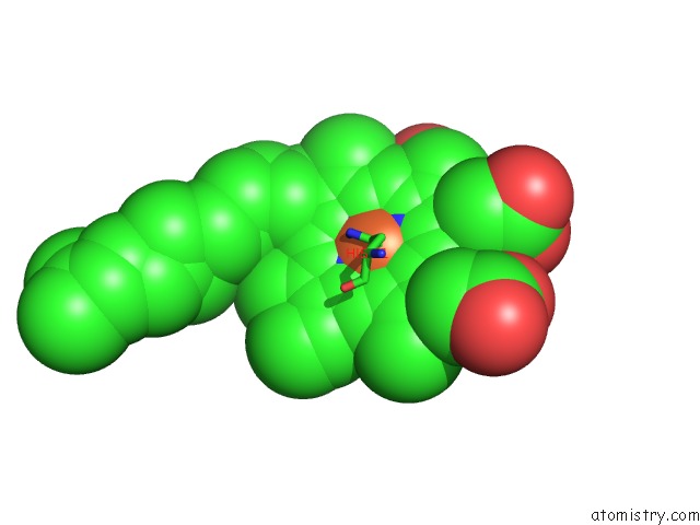
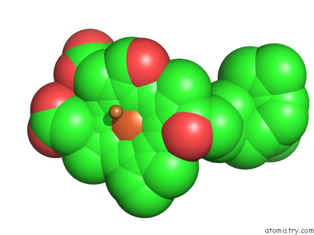
Iron Binding Sites:
The binding sites of Iron atom in the X-Ray Structure of Cyanide-Bound Bovine Heart Cytochrome C Oxidase in the Fully Oxidized State at 2.0 Angstrom Resolution
(pdb code 3x2q). This binding sites where shown within
5.0 Angstroms radius around Iron atom.
In total 4 binding sites of Iron where determined in the X-Ray Structure of Cyanide-Bound Bovine Heart Cytochrome C Oxidase in the Fully Oxidized State at 2.0 Angstrom Resolution, PDB code: 3x2q:
Jump to Iron binding site number: 1; 2; 3; 4;
In total 4 binding sites of Iron where determined in the X-Ray Structure of Cyanide-Bound Bovine Heart Cytochrome C Oxidase in the Fully Oxidized State at 2.0 Angstrom Resolution, PDB code: 3x2q:
Jump to Iron binding site number: 1; 2; 3; 4;
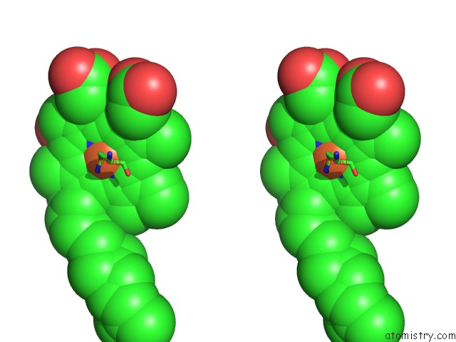
Iron binding site 1 out of 4 in 3x2q
Go back to
Iron binding site 1 out
of 4 in the X-Ray Structure of Cyanide-Bound Bovine Heart Cytochrome C Oxidase in the Fully Oxidized State at 2.0 Angstrom Resolution
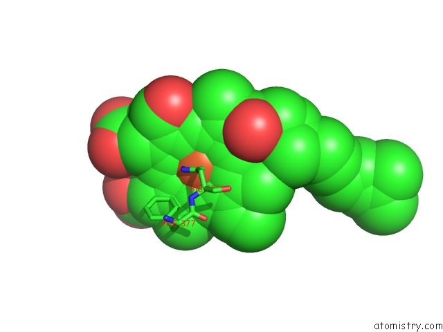
Mono view
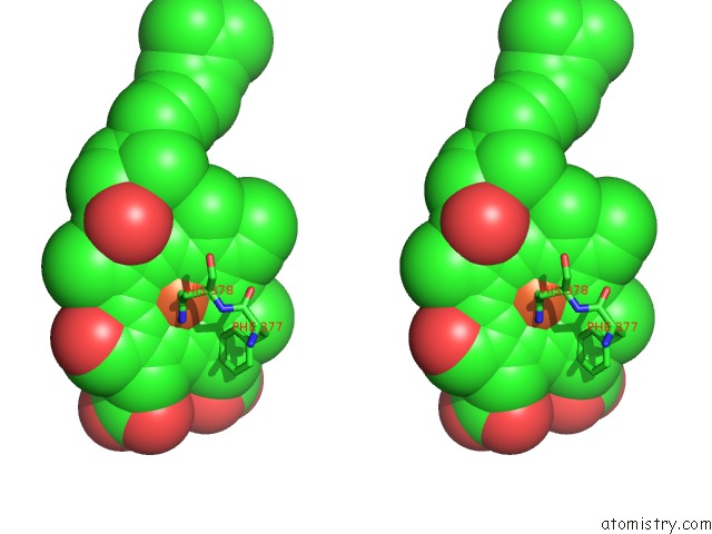
Stereo pair view
Mono view
Stereo pair view
A full contact list of Iron with other atoms in the Fe binding
site number 1 of X-Ray Structure of Cyanide-Bound Bovine Heart Cytochrome C Oxidase in the Fully Oxidized State at 2.0 Angstrom Resolution within 5.0Å range:
|
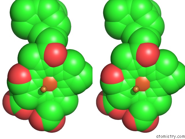
Iron binding site 2 out of 4 in 3x2q
Go back to
Iron binding site 2 out
of 4 in the X-Ray Structure of Cyanide-Bound Bovine Heart Cytochrome C Oxidase in the Fully Oxidized State at 2.0 Angstrom Resolution
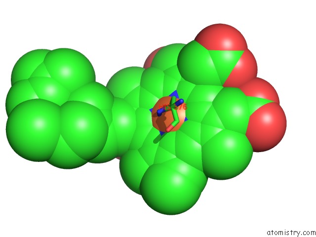
Mono view
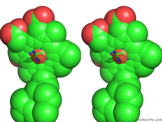
Stereo pair view
Mono view
Stereo pair view
A full contact list of Iron with other atoms in the Fe binding
site number 2 of X-Ray Structure of Cyanide-Bound Bovine Heart Cytochrome C Oxidase in the Fully Oxidized State at 2.0 Angstrom Resolution within 5.0Å range:
|
Iron binding site 3 out of 4 in 3x2q
Go back to
Iron binding site 3 out
of 4 in the X-Ray Structure of Cyanide-Bound Bovine Heart Cytochrome C Oxidase in the Fully Oxidized State at 2.0 Angstrom Resolution
Mono view
Stereo pair view
Mono view
Stereo pair view
A full contact list of Iron with other atoms in the Fe binding
site number 3 of X-Ray Structure of Cyanide-Bound Bovine Heart Cytochrome C Oxidase in the Fully Oxidized State at 2.0 Angstrom Resolution within 5.0Å range:
|
Iron binding site 4 out of 4 in 3x2q
Go back to
Iron binding site 4 out
of 4 in the X-Ray Structure of Cyanide-Bound Bovine Heart Cytochrome C Oxidase in the Fully Oxidized State at 2.0 Angstrom Resolution
Mono view
Stereo pair view
Mono view
Stereo pair view
A full contact list of Iron with other atoms in the Fe binding
site number 4 of X-Ray Structure of Cyanide-Bound Bovine Heart Cytochrome C Oxidase in the Fully Oxidized State at 2.0 Angstrom Resolution within 5.0Å range:
|
Reference:
N.Yano,
K.Muramoto,
M.Mochizuki,
K.Shinzawa-Itoh,
E.Yamashita,
S.Yoshikawa,
T.Tsukihara.
X-Ray Structure of Cyanide-Bound Bovine Heart Cytochrome C Oxidase in the Fully Oxidized State at 2.0 Angstrom Resolution. Acta Crystallogr F Struct V. 71 726 2015BIOL Commun.
ISSN: ESSN 2053-230X
PubMed: 26057802
DOI: 10.1107/S2053230X15007025
Page generated: Sun Aug 4 22:58:38 2024
ISSN: ESSN 2053-230X
PubMed: 26057802
DOI: 10.1107/S2053230X15007025
Last articles
F in 4OLHF in 4OKX
F in 4OKW
F in 4OKB
F in 4OKT
F in 4OK1
F in 4OJB
F in 4OJR
F in 4OI1
F in 4OJ9