Iron »
PDB 3x20-3zjo »
3ze7 »
Iron in PDB 3ze7: 3D Structure of the Ni-Fe-Se Hydrogenase From D. Vulgaris Hildenborough in the Reduced State at 1.95 Angstroms
Enzymatic activity of 3D Structure of the Ni-Fe-Se Hydrogenase From D. Vulgaris Hildenborough in the Reduced State at 1.95 Angstroms
All present enzymatic activity of 3D Structure of the Ni-Fe-Se Hydrogenase From D. Vulgaris Hildenborough in the Reduced State at 1.95 Angstroms:
1.12.7.2;
1.12.7.2;
Protein crystallography data
The structure of 3D Structure of the Ni-Fe-Se Hydrogenase From D. Vulgaris Hildenborough in the Reduced State at 1.95 Angstroms, PDB code: 3ze7
was solved by
M.C.Marques,
R.Coelho,
I.A.C.Pereira,
P.M.Matias,
with X-Ray Crystallography technique. A brief refinement statistics is given in the table below:
| Resolution Low / High (Å) | 45.78 / 1.95 |
| Space group | P 21 21 21 |
| Cell size a, b, c (Å), α, β, γ (°) | 72.297, 96.987, 103.855, 90.00, 90.00, 90.00 |
| R / Rfree (%) | 15.3 / 19 |
Other elements in 3ze7:
The structure of 3D Structure of the Ni-Fe-Se Hydrogenase From D. Vulgaris Hildenborough in the Reduced State at 1.95 Angstroms also contains other interesting chemical elements:
| Nickel | (Ni) | 1 atom |
Iron Binding Sites:
Pages:
>>> Page 1 <<< Page 2, Binding sites: 11 - 14;Binding sites:
The binding sites of Iron atom in the 3D Structure of the Ni-Fe-Se Hydrogenase From D. Vulgaris Hildenborough in the Reduced State at 1.95 Angstroms (pdb code 3ze7). This binding sites where shown within 5.0 Angstroms radius around Iron atom.In total 14 binding sites of Iron where determined in the 3D Structure of the Ni-Fe-Se Hydrogenase From D. Vulgaris Hildenborough in the Reduced State at 1.95 Angstroms, PDB code: 3ze7:
Jump to Iron binding site number: 1; 2; 3; 4; 5; 6; 7; 8; 9; 10;
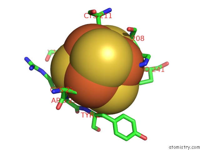
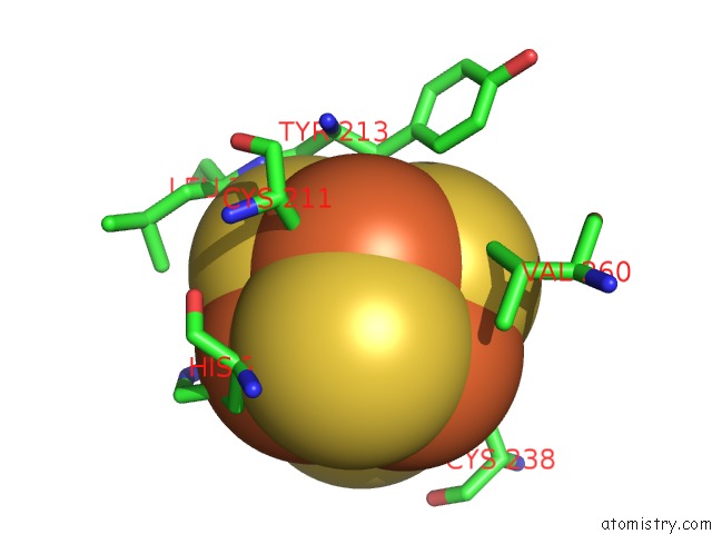
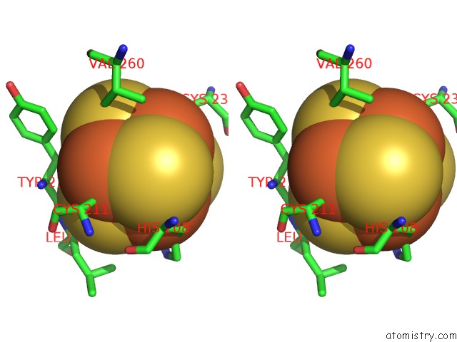
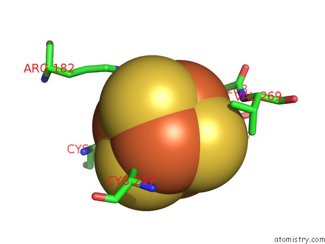
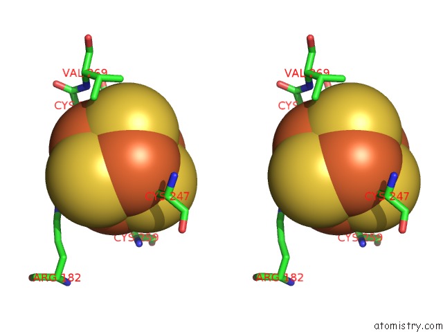
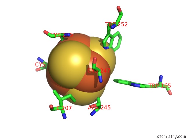
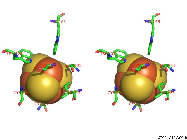
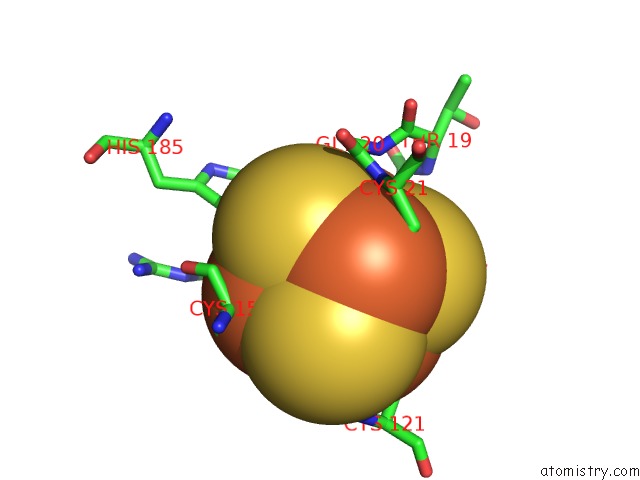
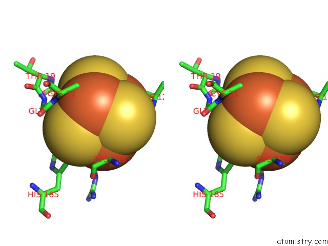
Iron binding site 1 out of 14 in 3ze7
Go back to
Iron binding site 1 out
of 14 in the 3D Structure of the Ni-Fe-Se Hydrogenase From D. Vulgaris Hildenborough in the Reduced State at 1.95 Angstroms
Mono view
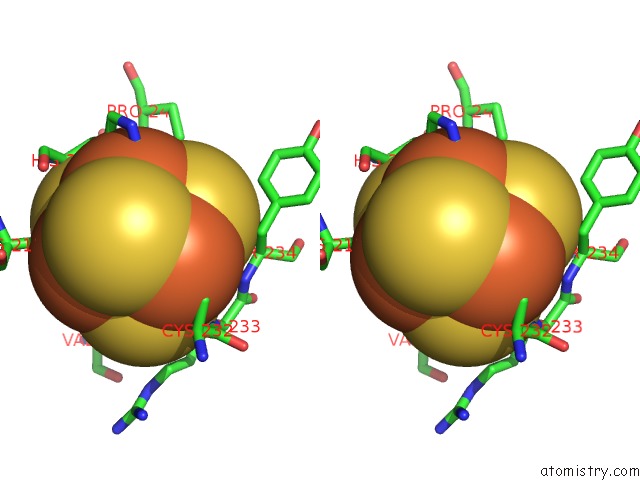
Stereo pair view
Mono view
Stereo pair view
A full contact list of Iron with other atoms in the Fe binding
site number 1 of 3D Structure of the Ni-Fe-Se Hydrogenase From D. Vulgaris Hildenborough in the Reduced State at 1.95 Angstroms within 5.0Å range:
|
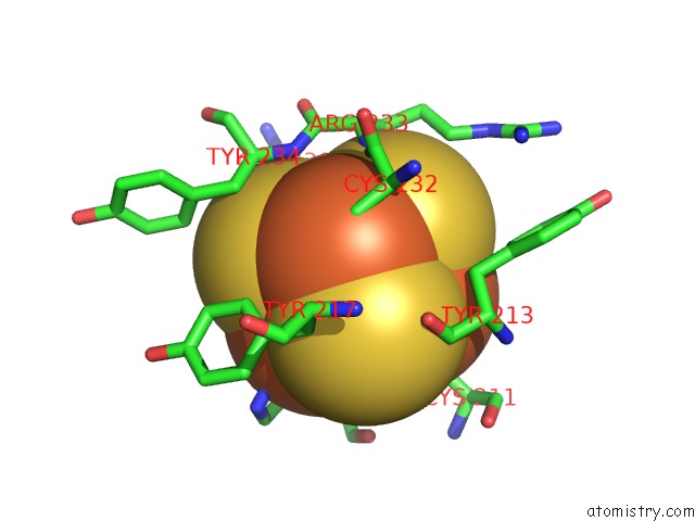
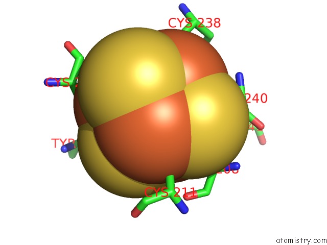
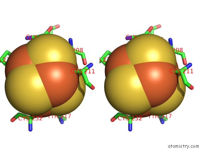
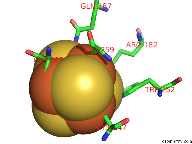
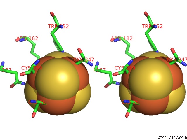
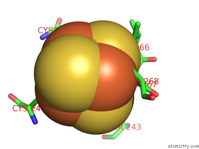
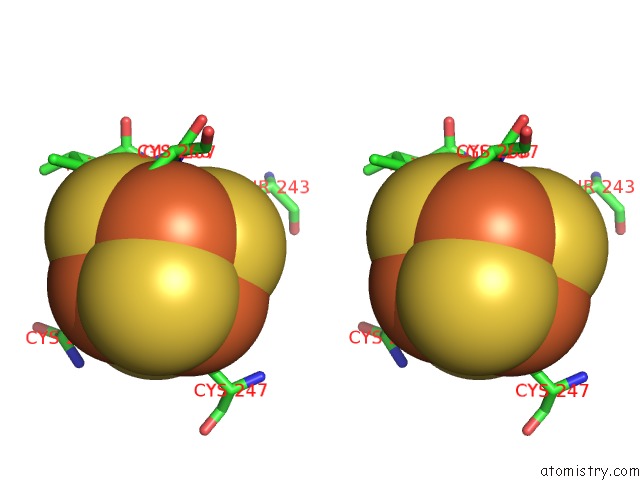
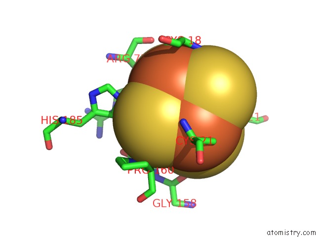
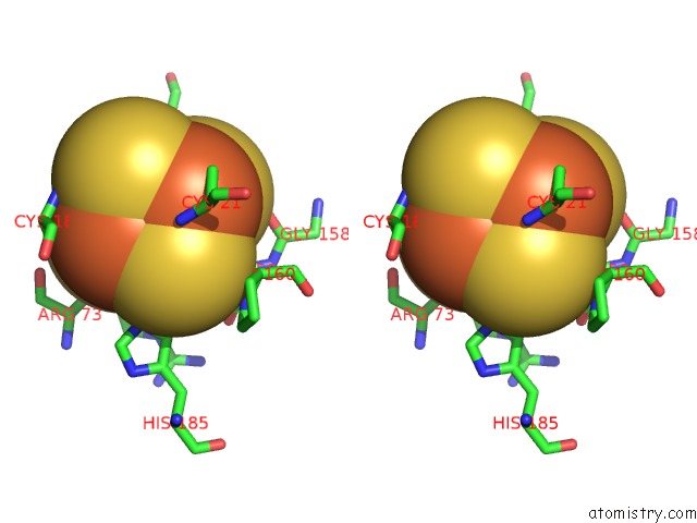
Iron binding site 2 out of 14 in 3ze7
Go back to
Iron binding site 2 out
of 14 in the 3D Structure of the Ni-Fe-Se Hydrogenase From D. Vulgaris Hildenborough in the Reduced State at 1.95 Angstroms
Mono view
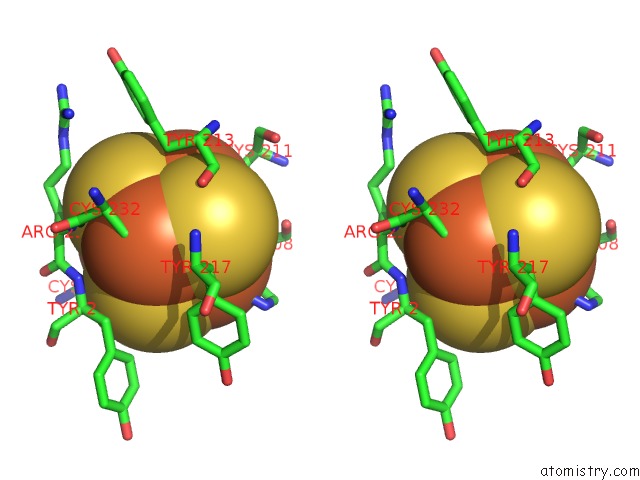
Stereo pair view
Mono view
Stereo pair view
A full contact list of Iron with other atoms in the Fe binding
site number 2 of 3D Structure of the Ni-Fe-Se Hydrogenase From D. Vulgaris Hildenborough in the Reduced State at 1.95 Angstroms within 5.0Å range:
|
Iron binding site 3 out of 14 in 3ze7
Go back to
Iron binding site 3 out
of 14 in the 3D Structure of the Ni-Fe-Se Hydrogenase From D. Vulgaris Hildenborough in the Reduced State at 1.95 Angstroms
Mono view
Stereo pair view
Mono view
Stereo pair view
A full contact list of Iron with other atoms in the Fe binding
site number 3 of 3D Structure of the Ni-Fe-Se Hydrogenase From D. Vulgaris Hildenborough in the Reduced State at 1.95 Angstroms within 5.0Å range:
|
Iron binding site 4 out of 14 in 3ze7
Go back to
Iron binding site 4 out
of 14 in the 3D Structure of the Ni-Fe-Se Hydrogenase From D. Vulgaris Hildenborough in the Reduced State at 1.95 Angstroms
Mono view
Stereo pair view
Mono view
Stereo pair view
A full contact list of Iron with other atoms in the Fe binding
site number 4 of 3D Structure of the Ni-Fe-Se Hydrogenase From D. Vulgaris Hildenborough in the Reduced State at 1.95 Angstroms within 5.0Å range:
|
Iron binding site 5 out of 14 in 3ze7
Go back to
Iron binding site 5 out
of 14 in the 3D Structure of the Ni-Fe-Se Hydrogenase From D. Vulgaris Hildenborough in the Reduced State at 1.95 Angstroms
Mono view
Stereo pair view
Mono view
Stereo pair view
A full contact list of Iron with other atoms in the Fe binding
site number 5 of 3D Structure of the Ni-Fe-Se Hydrogenase From D. Vulgaris Hildenborough in the Reduced State at 1.95 Angstroms within 5.0Å range:
|
Iron binding site 6 out of 14 in 3ze7
Go back to
Iron binding site 6 out
of 14 in the 3D Structure of the Ni-Fe-Se Hydrogenase From D. Vulgaris Hildenborough in the Reduced State at 1.95 Angstroms
Mono view
Stereo pair view
Mono view
Stereo pair view
A full contact list of Iron with other atoms in the Fe binding
site number 6 of 3D Structure of the Ni-Fe-Se Hydrogenase From D. Vulgaris Hildenborough in the Reduced State at 1.95 Angstroms within 5.0Å range:
|
Iron binding site 7 out of 14 in 3ze7
Go back to
Iron binding site 7 out
of 14 in the 3D Structure of the Ni-Fe-Se Hydrogenase From D. Vulgaris Hildenborough in the Reduced State at 1.95 Angstroms
Mono view
Stereo pair view
Mono view
Stereo pair view
A full contact list of Iron with other atoms in the Fe binding
site number 7 of 3D Structure of the Ni-Fe-Se Hydrogenase From D. Vulgaris Hildenborough in the Reduced State at 1.95 Angstroms within 5.0Å range:
|
Iron binding site 8 out of 14 in 3ze7
Go back to
Iron binding site 8 out
of 14 in the 3D Structure of the Ni-Fe-Se Hydrogenase From D. Vulgaris Hildenborough in the Reduced State at 1.95 Angstroms
Mono view
Stereo pair view
Mono view
Stereo pair view
A full contact list of Iron with other atoms in the Fe binding
site number 8 of 3D Structure of the Ni-Fe-Se Hydrogenase From D. Vulgaris Hildenborough in the Reduced State at 1.95 Angstroms within 5.0Å range:
|
Iron binding site 9 out of 14 in 3ze7
Go back to
Iron binding site 9 out
of 14 in the 3D Structure of the Ni-Fe-Se Hydrogenase From D. Vulgaris Hildenborough in the Reduced State at 1.95 Angstroms
Mono view
Stereo pair view
Mono view
Stereo pair view
A full contact list of Iron with other atoms in the Fe binding
site number 9 of 3D Structure of the Ni-Fe-Se Hydrogenase From D. Vulgaris Hildenborough in the Reduced State at 1.95 Angstroms within 5.0Å range:
|
Iron binding site 10 out of 14 in 3ze7
Go back to
Iron binding site 10 out
of 14 in the 3D Structure of the Ni-Fe-Se Hydrogenase From D. Vulgaris Hildenborough in the Reduced State at 1.95 Angstroms
Mono view
Stereo pair view
Mono view
Stereo pair view
A full contact list of Iron with other atoms in the Fe binding
site number 10 of 3D Structure of the Ni-Fe-Se Hydrogenase From D. Vulgaris Hildenborough in the Reduced State at 1.95 Angstroms within 5.0Å range:
|
Reference:
M.C.Marques,
R.Coelho,
I.A.C.Pereira,
P.M.Matias.
Redox State-Dependent Changes in the Crystal Structure of [Nifese] Hydrogenase From Desulfovibrio Vulgaris Hildenborough Int.J.Hydrogen Energy V. 38 8664 2013.
ISSN: ISSN 0360-3199
DOI: 10.1016/J.IJHYDENE.2013.04.132
Page generated: Tue Aug 5 08:34:50 2025
ISSN: ISSN 0360-3199
DOI: 10.1016/J.IJHYDENE.2013.04.132
Last articles
Fe in 4W7LFe in 4W7J
Fe in 4W7K
Fe in 4VHB
Fe in 4V3Z
Fe in 4V3Y
Fe in 4V1Y
Fe in 4V3X
Fe in 4V3W
Fe in 4V3V