Iron »
PDB 3zjp-4a7m »
3zkp »
Iron in PDB 3zkp: Structure of A Mutant of P450 Eryk in Complex with Erythromycin B.
Enzymatic activity of Structure of A Mutant of P450 Eryk in Complex with Erythromycin B.
All present enzymatic activity of Structure of A Mutant of P450 Eryk in Complex with Erythromycin B.:
1.14.13.154;
1.14.13.154;
Protein crystallography data
The structure of Structure of A Mutant of P450 Eryk in Complex with Erythromycin B., PDB code: 3zkp
was solved by
L.C.Montemiglio,
B.Vallone,
C.Savino,
with X-Ray Crystallography technique. A brief refinement statistics is given in the table below:
| Resolution Low / High (Å) | 29.12 / 2.00 |
| Space group | P 1 21 1 |
| Cell size a, b, c (Å), α, β, γ (°) | 57.753, 36.597, 96.273, 90.00, 92.93, 90.00 |
| R / Rfree (%) | 16.332 / 21.529 |
Iron Binding Sites:
The binding sites of Iron atom in the Structure of A Mutant of P450 Eryk in Complex with Erythromycin B.
(pdb code 3zkp). This binding sites where shown within
5.0 Angstroms radius around Iron atom.
In total only one binding site of Iron was determined in the Structure of A Mutant of P450 Eryk in Complex with Erythromycin B., PDB code: 3zkp:
In total only one binding site of Iron was determined in the Structure of A Mutant of P450 Eryk in Complex with Erythromycin B., PDB code: 3zkp:
Iron binding site 1 out of 1 in 3zkp
Go back to
Iron binding site 1 out
of 1 in the Structure of A Mutant of P450 Eryk in Complex with Erythromycin B.

Mono view
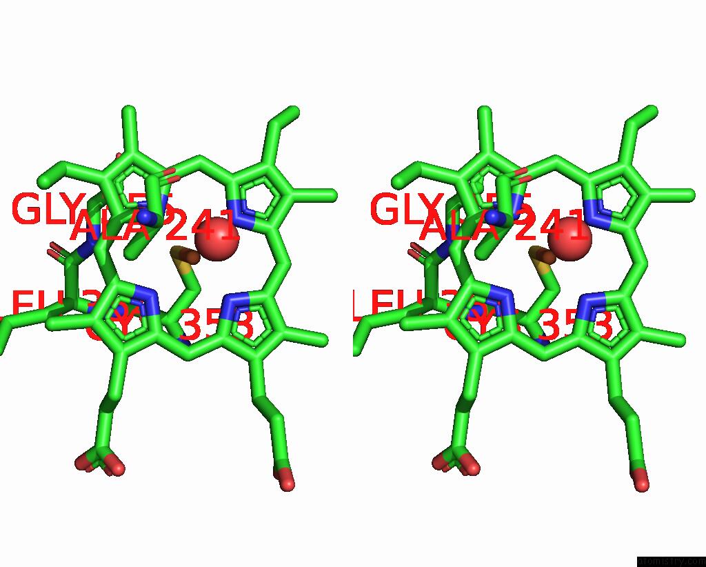
Stereo pair view
Mono view
Stereo pair view
A full contact list of Iron with other atoms in the Fe binding
site number 1 of Structure of A Mutant of P450 Eryk in Complex with Erythromycin B. within 5.0Å range:
|
Reference:
L.C.Montemiglio,
A.Macone,
C.Ardiccioni,
G.Avella,
B.Vallone,
C.Savino.
Redirecting P450 Eryk Specificity By Rational Site-Directed Mutagenesis. Biochemistry V. 52 3678 2013.
ISSN: ISSN 0006-2960
PubMed: 23597312
DOI: 10.1021/BI400223J
Page generated: Sun Aug 4 23:22:01 2024
ISSN: ISSN 0006-2960
PubMed: 23597312
DOI: 10.1021/BI400223J
Last articles
Zn in 9J0NZn in 9J0O
Zn in 9J0P
Zn in 9FJX
Zn in 9EKB
Zn in 9C0F
Zn in 9CAH
Zn in 9CH0
Zn in 9CH3
Zn in 9CH1