Iron »
PDB 3zjp-4a7m »
3zoi »
Iron in PDB 3zoi: Isopenicillin N Synthase with Ac-O-Methyl-D-Threonine
Enzymatic activity of Isopenicillin N Synthase with Ac-O-Methyl-D-Threonine
All present enzymatic activity of Isopenicillin N Synthase with Ac-O-Methyl-D-Threonine:
1.21.3.1;
1.21.3.1;
Protein crystallography data
The structure of Isopenicillin N Synthase with Ac-O-Methyl-D-Threonine, PDB code: 3zoi
was solved by
P.J.Rutledge,
I.J.Clifton,
W.Ge,
with X-Ray Crystallography technique. A brief refinement statistics is given in the table below:
| Resolution Low / High (Å) | 11.96 / 1.82 |
| Space group | P 21 21 21 |
| Cell size a, b, c (Å), α, β, γ (°) | 46.480, 70.790, 100.510, 90.00, 90.00, 90.00 |
| R / Rfree (%) | 16 / 20.2 |
Iron Binding Sites:
The binding sites of Iron atom in the Isopenicillin N Synthase with Ac-O-Methyl-D-Threonine
(pdb code 3zoi). This binding sites where shown within
5.0 Angstroms radius around Iron atom.
In total only one binding site of Iron was determined in the Isopenicillin N Synthase with Ac-O-Methyl-D-Threonine, PDB code: 3zoi:
In total only one binding site of Iron was determined in the Isopenicillin N Synthase with Ac-O-Methyl-D-Threonine, PDB code: 3zoi:
Iron binding site 1 out of 1 in 3zoi
Go back to
Iron binding site 1 out
of 1 in the Isopenicillin N Synthase with Ac-O-Methyl-D-Threonine
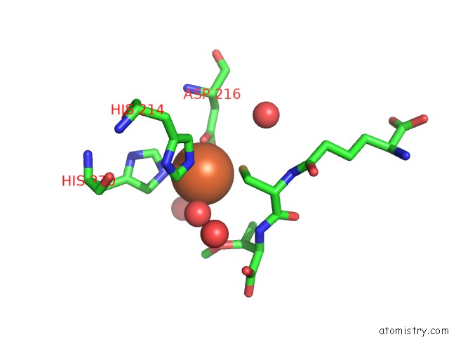
Mono view
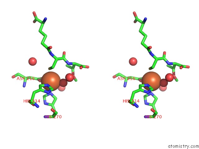
Stereo pair view
Mono view
Stereo pair view
A full contact list of Iron with other atoms in the Fe binding
site number 1 of Isopenicillin N Synthase with Ac-O-Methyl-D-Threonine within 5.0Å range:
|
Reference:
I.J.Clifton,
W.Ge,
R.M.Adlington,
J.E.Baldwin,
P.J.Rutledge.
The Crystal Structure of An Isopenicillin N Synthase Complex with An Ethereal Substrate Analogue Reveals Water in the Oxygen Binding Site. Febs Lett. V. 587 2705 2013.
ISSN: ISSN 0014-5793
PubMed: 23860486
DOI: 10.1016/J.FEBSLET.2013.07.016
Page generated: Sun Aug 4 23:23:13 2024
ISSN: ISSN 0014-5793
PubMed: 23860486
DOI: 10.1016/J.FEBSLET.2013.07.016
Last articles
Zn in 9J0NZn in 9J0O
Zn in 9J0P
Zn in 9FJX
Zn in 9EKB
Zn in 9C0F
Zn in 9CAH
Zn in 9CH0
Zn in 9CH3
Zn in 9CH1